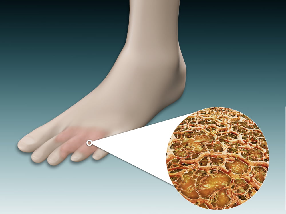
Anatomy of foot fungus with microscopic closeup Poster Print (16 x 12)
Before giving you the diagnosis, your dermatologist may also take some samples. Collecting a bit of debris from beneath a nail, trimming off part your nail, or scraping off a bit of skin can be very helpful. In a lab, these samples can be examined under a microscope to find out what's causing the problem.
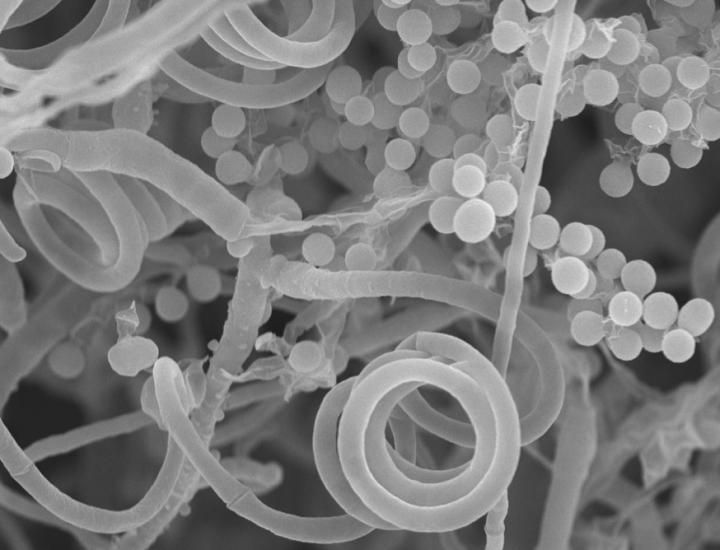
Toenail Fungus' Nonexistent Sex Life Is More Interesting Than You Think Live Science
If necessary, mycological (a branch of biology that deals with fungus) cure can be determined with a negative potassium hydroxide (KOH) preparation (the sample is examined under a microscope) and negative fungal culture (a sample is given time to grow to see if fungus is present). Available Treatments
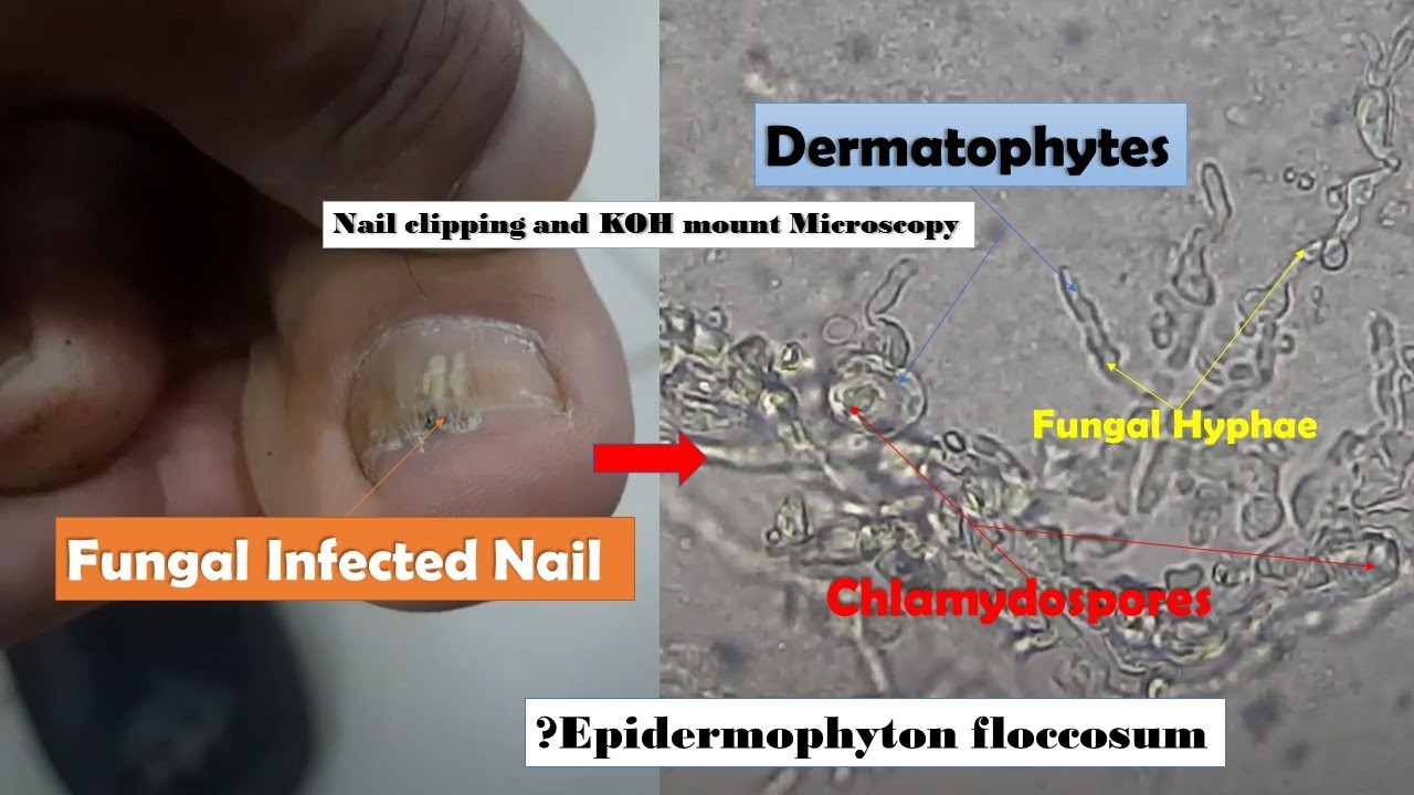
Fungal Infected Nail Microscopy showing Hyphae and Chlamydospores of Dermatophytes YouTube
To confirm the diagnosis, the healthcare provider might collect a nail clipping to look at under a microscope or to send to a laboratory for testing. Treatment Fungal nail infections can be difficult to cure, and treatment is most successful when started early.
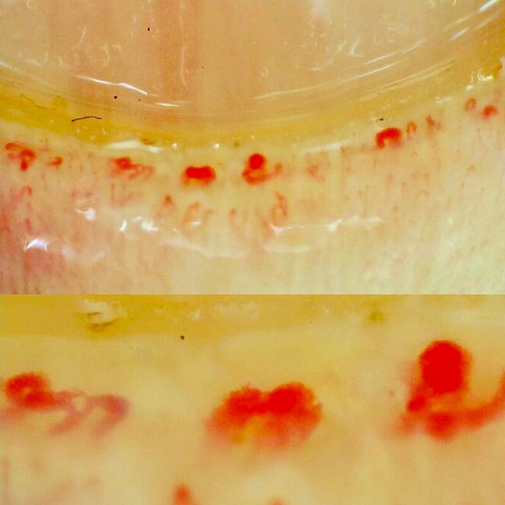
Toenail Microscope capillaroscopy,nailfold capillaroscopy,fingernail under microscope,nail
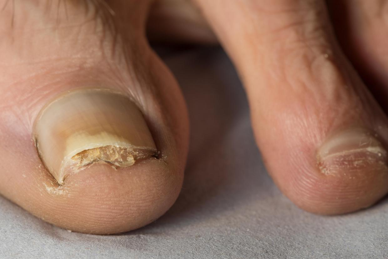
Causes Symptoms Diagnosis Treatment Onychomycosis is a fungal infection of the nails. (See also Overview of Nail Disorders .) About 10% of people have onychomycosis, which most often affects the toenails rather than the fingernails.
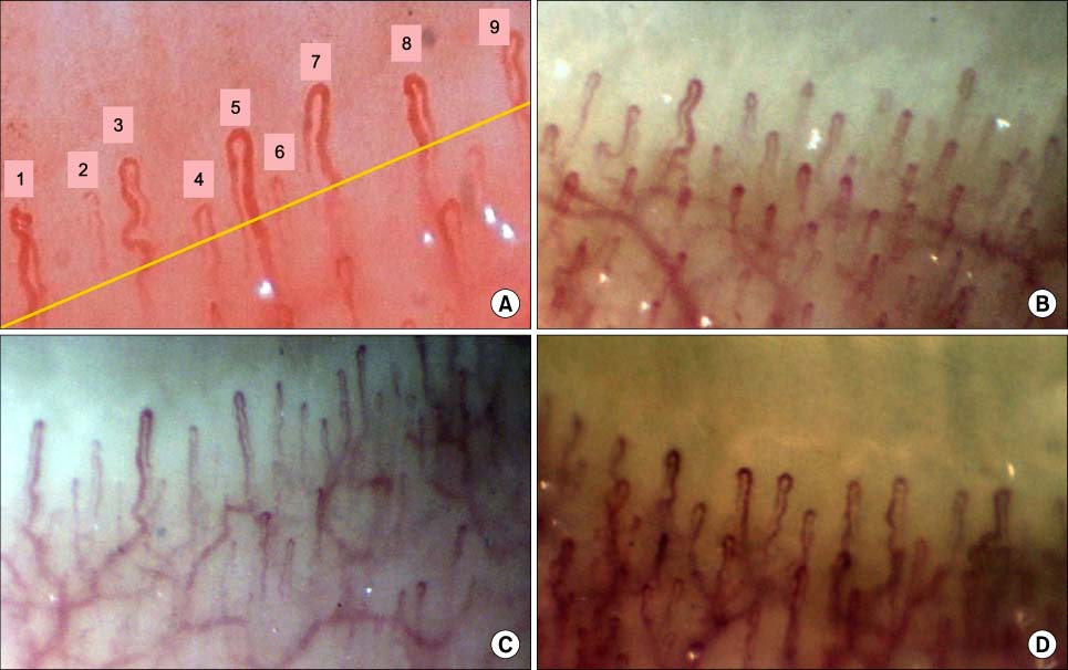
Toenail Microscope capillaroscopy,nailfold capillaroscopy,fingernail under microscope,nail
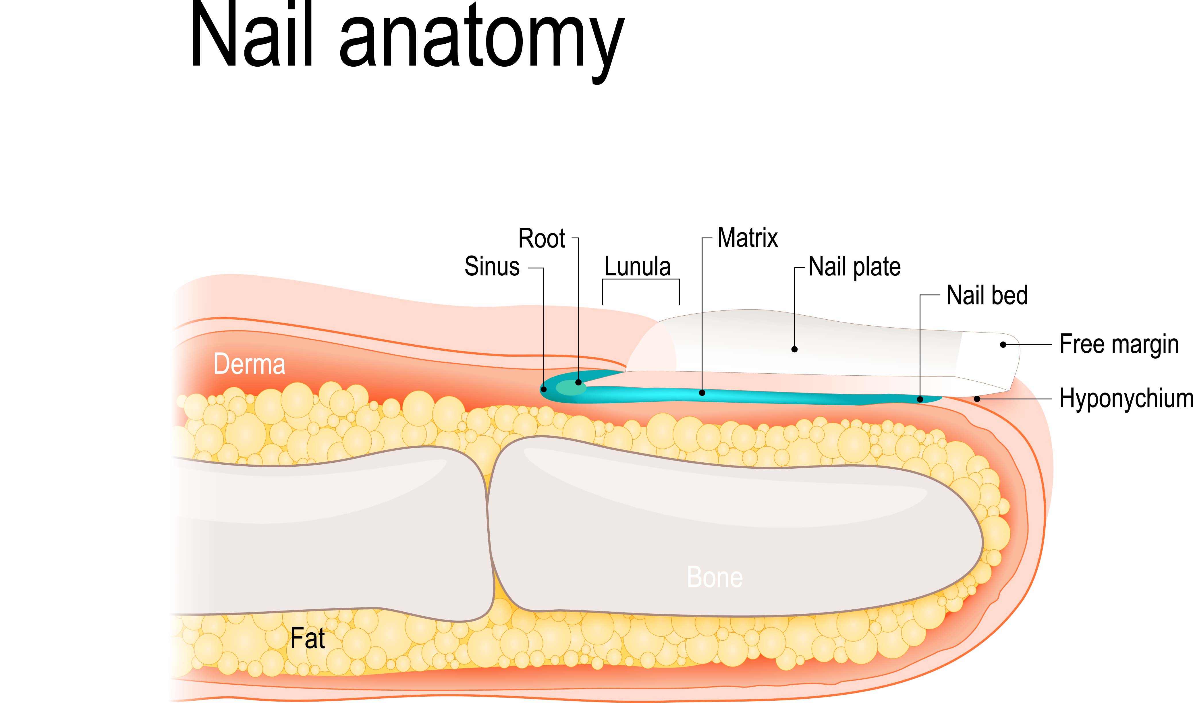
Nails Under The Microscope - Understanding Nail Anatomy & Disorders by Richard K. Scher, MD | June 1, 1996 | While a nail technician's job is focused primarily on the exterior surfaces of the nail, the supporting structures are just as important, as they dictate how the nails grow.

Scanning electron microscopy of the nail plate in onychomycosis patients with negative fungal
A sample of the affected nail must be examined under a microscope to confirm if fungi are present, and which type. This information can help the doctor determine how to treat the infection, if at all," says Dr. Milliman. For many people, nail fungus is primarily a cosmetic concern.
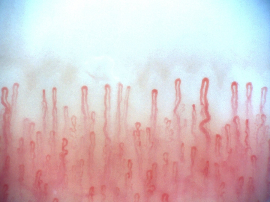
Toenail Microscope capillaroscopy,nailfold capillaroscopy,fingernail under microscope,nail
These considerations may warrant antifungal treatment in the absence of hyphae under the microscope. 2 In a European study of 45,000 patients with suspected onychomycosis, general physicians.

View of a subungual haematoma under a toenail Stock Image M330/0754 Science Photo Library
According to Dr. Lipner, doctors usually confirm toenail fungus by examining a clipping under a microscope.. If there's fungus under multiple nails, or if the toenails are extra thick, Dr.

Infected nail, SEM Stock Image B220/1415 Science Photo Library
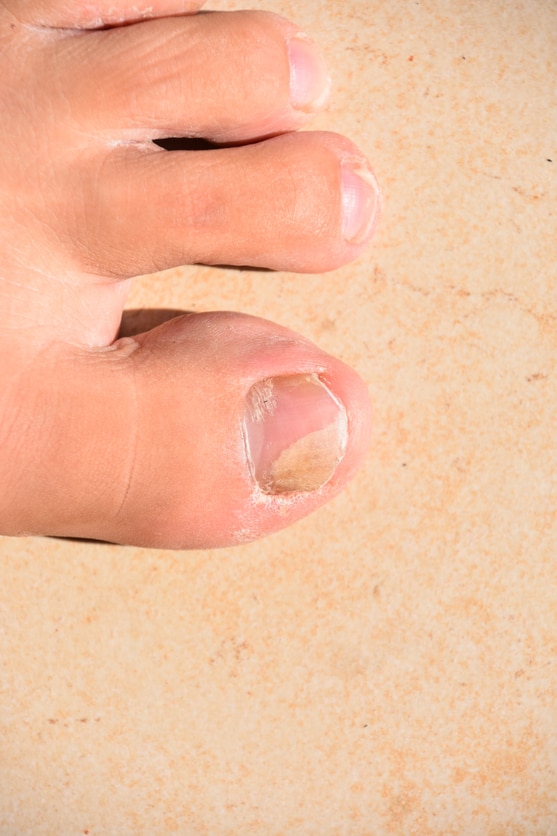
Physical Characteristics of Infected Toenails Identifying toenail fungus goes beyond recognizing visible symptoms. Under the microscope, the physical characteristics of infected toenails reveal intricate details about the severity and type of infection.
Ingrown Toenail Surgery Part A The Anatomy Elizabeth E. Auger, DPM
Onychoscopy is a new method that can help the physician, as in onychomycosis, it shows a typical fringed proximal margin. Treatment is chosen depending on the modality of nail invasion, fungus species and the number of affected nails. Oral treatments are often limited by drug interactions, while topical antifungal lacquers have less efficacy.
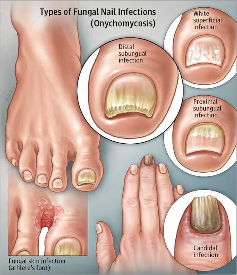
White Superficial Onychomycosis Types, Symptoms, Causes & Treatment
This video shows how a nail fungus look like under microscope. It is always good to know things in close up. The video contains below details.1.Description o.
Fungal nail infection nidirect
The diagnosis can be confirmed by looking at scrapings from the nail under a microscope. This can help determine the type of fungus. Samples can also be sent to a lab for a culture.
Pictures of Toenail Fungus Is This What You Have?
Fungal infection of the toenails or fingernails is a superficial fungus infection (dermatophytosis). The infection is caused by a fungal microbe that invades the nail bed. Fungal nail infection is also termed onychomycosis and tinea unguium.

Toenail Under Microscope Query Mores
This is what nail fungus looks like close up under a 2000x microscope. It was hard to keep the finger still so the focus kept changing. I tried digging the f.
How to Tell if You Have a Toenail Fungus Colorado Center of Orthopaedic Excellence
Dermatophyte nail infections are commonly known as dermatophyte onychomycosis or tinea unguium.. including direct examination under a microscope, Wood's light examination, or fungal cultures. Treatment of dermatophytosis depends on the infectious microorganism, location and severity of the infection, and typically includes systemic or.
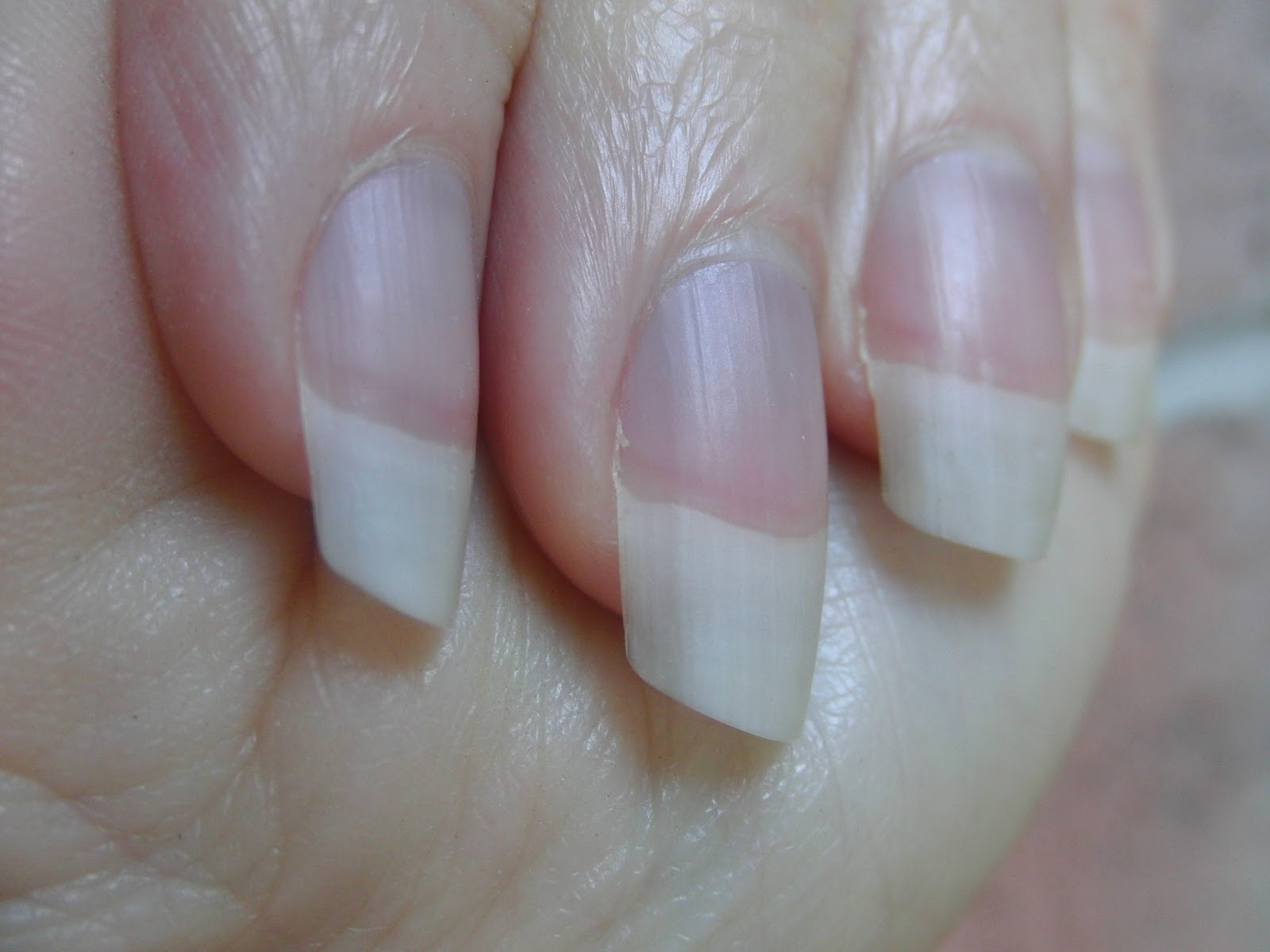
Beauty Blog by Lisa TIPS from head to toe! The structure of your nail
Discover toenail fungus secrets under a microscope. Act now and visit fungusless.co. #fungusless #toenailfungus #he.
