
liver lobes anatomy Liver anatomy, Bile duct, Abdomen
Introduction. Riedel's liver lobe, the most well-known accessory lobe of the liver, is considered a rare anatomical variant of the liver morphology with a downward tongue-like projection of the anterior edge of the right lobe of the liver involving the segments V and VI [1,2].It is usually detected incidentally with abdominal imaging performed for other indications and is asymptomatic.

Liver Basicmedical Key
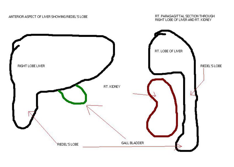
A 46-year-old woman patient presented with a lump right hypochondrium with a diagnosis of calculus cholecystitis on ultrasound abdomen. During laparoscopy, a Riedel's lobe of liver was seen (Fig. 1).Riedel lobe is a tongue-like, inferior projection of the right lobe of the liver beyond the level of the most inferior costal cartilage, and it is an anatomical variant.

Riedel lobe Radiology Reference Article Radiology, Lobes, Riedel
Riedel's lobe may present with minor symptoms like abdominal discomfort, nausea, constipation, and vomiting. This discomfort is caused due to compression caused extrinsically or there can be no cause. The symptoms of Riedel's liver be the same as any of the following conditions: Emphysema. Congestive heart failure.
A Gallery of HighResolution, Ultrasound, Color Doppler & 3D Images Liver
Preoperative imaging showed a downward elongation of the liver, ending at the level of the iliac crest. Originally reported in 1888 by a German Surgeon named Carl Ludwig Riedel in seven female patients who had palpable masses, they were all confirmed by surgical exploration. The potential complications are rare and include torsion, mass effect.

Riedel’s lobe of liver
Riedel's liver lobe, the most well-known accessory lobe of the liver, is considered a rare anatomical variant of the liver morphology with a downward tongue-like projection of the anterior edge of.
Cureus Coexistence of Riedel’s Lobe and Supernumerary Kidney as Random Imaging Findings
Riedel's lobe is a normal variant form of right liver lobe rarely found. Here we report a case of 38 years old female with an incidental finding not revealed in physical examination, but then known to have hepatomegaly by gynecological ultrasonography. Diagnosis of Riedel's lobe was strengthened by.

PPT Liver & Spleen PowerPoint Presentation, free download ID2118838
Introduction. Riedel's liver lobe, the most well-known accessory lobe of the liver, is considered a rare anatomical variant of the liver morphology with a downward tongue-like projection of the anterior edge of the right lobe of the liver involving the segments V and VI .It is usually detected incidentally with abdominal imaging performed for other indications and is asymptomatic.

riedel's lobe
Riedel's liver lobe, the most well-known accessory lobe of the liver, is considered a rare anatomical variant of the liver morphology with a downward tongue-like projection of the anterior edge of.

Morphological variations of the liver and its applied significance A cadaveric study Sangeetha
Riedel's lobe appears to be a common variant of normal anatomy, its prevalence being dependent on age-related changes in liver size and skeletal shape. Riedel's lobe should be considered in all patients undergoing cross-sectional imaging. It may harbor a lesion that might not be demonstrated unless the most inferior aspect of the liver is imaged.
Riedel Lobe Of Liver On Ultrasound Https Encrypted Tbn0 Gstatic Com Images Q Tbn
On exploratory laparotomy a 10x6cm-sized tongue-like lobe from left-lobe of liver was seen extending to the umbilicus and compressing the prepyloric area of the stomach [6]. The presence of Riedel's lobe can be challenging for the nephrologist performing laproscopic interventions [5, 13]. Riedel's lobe chiefly presents two special technical.
Riedel's Lobe Location, Symptoms, Complications, Surgery
Beaver tail liver is an elongated left liver lobe that extends laterally to contact and surround the spleen ( normal variant ). This is also known as a sliver of liver and is a variant of hepatic morphology. This variant is more common in females. The liver parenchyma is normal and thereby has the same risks of hepatic pathology as the rest of.

Riedel lobe Image
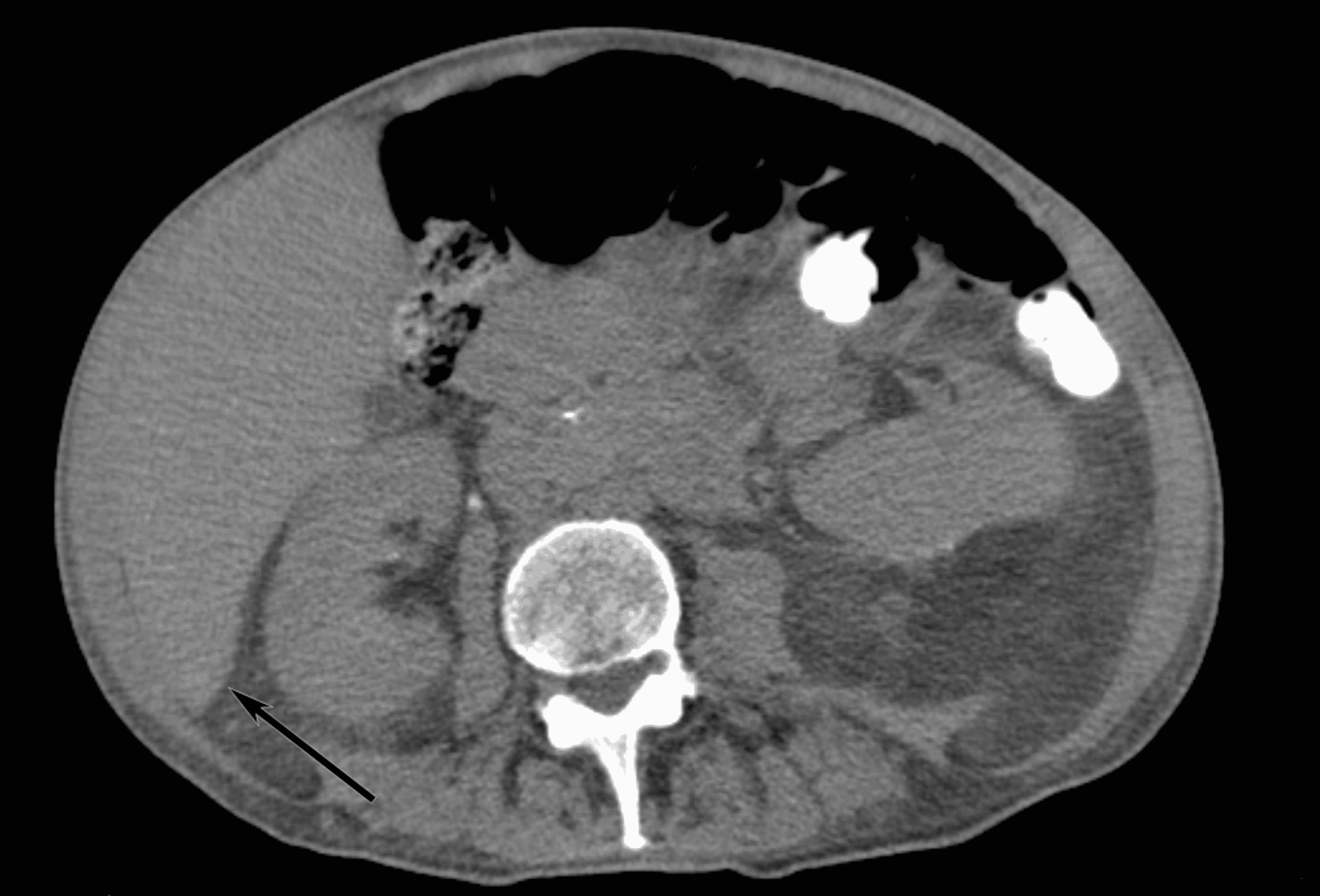
The kidney is in its normal position in the retroperitoneum and is not displaced downward. This suggests that the liver enlargement is from a Riedel lobe, a normal variant of the liver with a large right hepatic lobe. With hepatomegaly, the entire liver would be enlarged and displace intra-abdominal organs.

Riedel’s lobe of liver
Riedel lobe is a common anatomical variant of the liver to be aware of because it can simulate a mass. Its misidentification as a pathologic abdominal mass has led to surgery. Pathology can also occur within it (e.g. malignancy or even torsion) a.

Riedel\'s Lobe Did you know these facts? Eurorad
Riedel lobe of the liver is a simple anatomical variation, a downward tongue-like projection of the anterior edge of the right lobe of the liver to the right of the gallbladder with its typical case to be rare. We report the case of a 71-year-old woman with typical feature of a nonpalpable Riedel's lobe of the liver, as an incidental finding.

Riedels Lobe Liver Stock Illustration 1777418417 Shutterstock
Riedel's lobe is an anatomical variation, and the exact etiology is still unclear. 1, 2 The reported incidence varies considerably (3.3-31%); this may be because the diagnostic criteria and methods are different from the previous reports. 1, 2, 3 The original definition of Riedel's lobe is a downward tongue-like projection of the right.

PPT What is The LIVER ? PowerPoint Presentation ID373722
Accessory liver lobes are defined as morphologic variations of the liver and are related to excessive development [1], [2]. The presence of an accessory liver lobe is often revealed by torsion, especially when the lobe is pedunculated [3]. The diagnosis is usually made by imaging. Riedel's lobe is the most well-known of accessory liver lobes [4.
