Hematoxylin and Eosin (H&E) Stain Lab Tests Guide
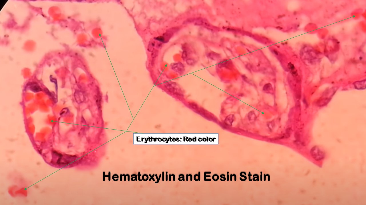
Hematoxylin-eosin staining is based on the affinity of two dyes, hematoxylin and eosin, to tissue components and structures, due to their pH value. Hematoxylin is a basic "nuclear" dye which has affinities for acidic structures, such as DNA, RNA, cell membrane proteins, and elastin.

Hematoxylin and eosin staining demonstrating as follows. (a) CCRCC... Download Scientific Diagram
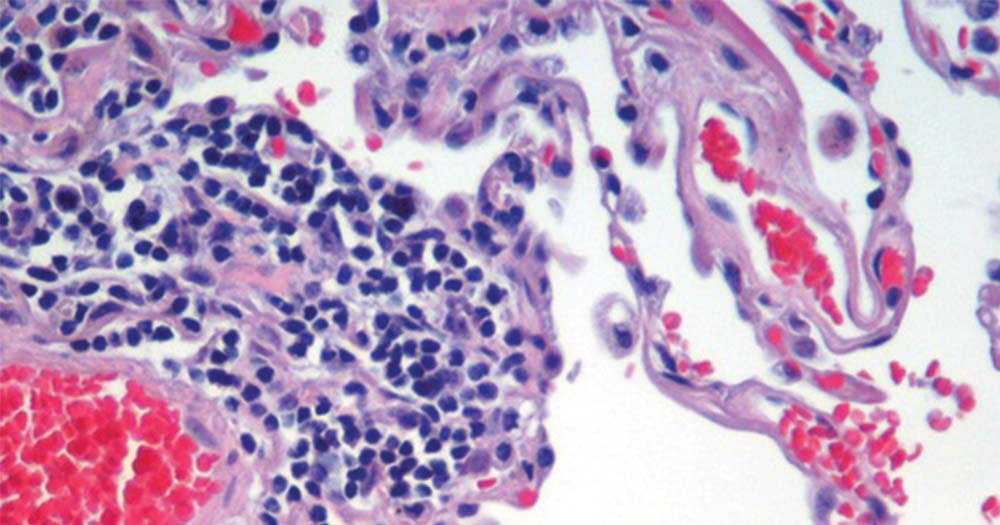
Hematoxylin and eosin staining is among the oldest and most common techniques in histology. The two-component dye differentially stains tissue components. Hematoxylin with a mordant is positively charged and stains nucleic acid and ribosomes blue. Eosin is negatively charged and stains proteins, such as collagen and elastin pink.

Hematoxylin and eosin staining (H&E) of infiltrated leukocytes (IL;... Download Scientific Diagram
Hematoxylin and eosin (H&E) are the principal stains applied for the demonstration of the nucleus and the cytoplasmic inclusions. Harri's hematoxylin (primary stain) contains alum and alum acts as a mordant that stains the nucleus light blue which turns red in the presence of acid.
Hematoxylin and Eosin Stain Introduction, Principle, Procedure, Result
Hematoxylin and Eosin (H&E) staining is a widely used histological staining technique in pathology and histology. It is used to colourize tissues and cellular structures to observe and study them under a microscope. STAINING Staining is a process by which a colour is imparted to sectioned tissue.

(A) Hematoxylin and eosinstained section (Original magnification... Download Scientific Diagram
Stain Rinse: Remove excess stain by rinsing in running tap water for 1+ minutes. Destain and Differentiate: This is the critical step that determines the quality of the final preparation. Destain in 0.3% acid alcohol until the cytoplasm has only a faint stain but the sharp nuclear stain still remains. 3 or 4 dips.
Hematoxylin and Eosin (H&E) Staining Principle, Procedure and Interpretation
Hematoxylin, generally without eosin, is useful as a counterstain for many immunohistochemical or hybridization procedures that use colorimetric substrates (such as alkaline phosphatase or peroxidase). This protocol describes H&E staining of tissue and cell sections.

Hematoxylineosin staining image of chronic group testis tissue. a... Download Scientific Diagram
The H&E staining method involves application of haematoxylin mixed with a metallic salt, or mordant, often followed by a rinse in a weak acid solution to remove excess staining ( differentiation ), followed by bluing in mildly alkaline water.

Representative histology with H&E (hematoxylin and eosin staining) at... Download Scientific
The principle behind H & E stain is the chemical attraction between tissue and dye. Hematoxylin, a basic dye imparts blue-purple contrast on basophilic structures, primarily those containing nucleic acid moeties such as chromtatin, ribosomes and cytoplasmic regions rich in RNA.

(A) Representative photomicrographs of hematoxylin and eosine staining... Download Scientific
HE Staining: Principle, Procedure, and Interpretation | Haematoxylin and Eosin Staining |Welcome to our comprehensive guide on HE staining (Hematoxylin and E.
Check out Hematoxylin And Eosin Staining Protocol Laboratory Hub
Hematoxylin and Eosin (H&E) Staining Protocol. The oxidation product of haematoxylin is haematin, and is the active ingredient in the staining solution. Haematoxylin is not classified as a dye since the molecule possesses no chromophore. The in situ oxidation of haematoxylin is effected by the addition of a strong oxidant to the stain, in this.

Hematoxylin and eosin (H&E) staining (left image of each timepoint and... Download Scientific
The staining procedure for H&E follows a basic protocol: Dewaxing Dehydration Hematoxylin Differentiation Bluing Eosin Dehydration Clearing Cover-slipping The format is easily reproduced and the reagents resilient enough to allow for large numbers of slides to be stained consistently before reagents need to be changed.

Hematoxylin & Eosin (H&E) staining and Masson staining on kidney tissue... Download Scientific
Protocol for H&E staining: While sections are in water, skim surface of hematoxalin with a Kimwipe to remove oxidized particles. Blot excess water from slide holder before going into hematoxalin. Coverslip slides using Permount (xylene based). Place a drop of Permount on the slide using a glass rod, taking care to leave no bubbles.

Hematoxylin and eosin (H&E) staining (left image of each timepoint and... Download Scientific
Histological staining: hematoxylin & eosin. The most popular and one of the principal stains in histology is hematoxylin and eosin stain. It gives us an overview of the tissue and its structure.. 03:39 5.01 MB 108,505.

Hematoxylin and eosinstained histologic sections at 20Â magnification... Download Scientific
Stains may be used to define & examine the bulk tissues, Cell population or Organelles within the individual cells. In histopathology laboratory, the Hematoxylin and Eosin stain is referred as the Routine Stain as it can be used to stain any tissue specimen to reveal the underlying tissue structures and conditions.
(AF) Hematoxylin and eosin (HE) staining (A), and immunohistochemistry... Download Scientific
Hematoxylin and Eosin (H&E) staining is a commonly used histological staining technique that is used to visualize the structure of cells and tissues in a sample. The staining is performed by first staining the tissue with hematoxylin, a basic dye that stains acidic structures such as the cytoplasm and nuclei blue.

Hematoxylineosin staining mass composed of lymphoplasmocytic... Download Scientific Diagram
H & E stain has the following steps in the procedure. Place the section fixed tissue slid in the xylene for 3 minutes. In the next step, transfer it to the absolute alcohol for 3 minutes. Place it in the methylated spirit for two to three minutes.
