
Artery and Vein Cross Section Diagram Quizlet
Cross section Cross-section The chambers of the heart operate as a 'double-pump' system for the body's circulation. In coordination with valves, the chambers work to keep blood flowing in the.
Coronary artery cross section Stock Vector Images Alamy
Currently, coronary artery cross-sectional areas are easily measured with MDCT and potential narrowing degrees can be defined in a more objective manner. Therefore, we formulated the correlation between cross-sectional areas of coronary arteries instead of diameters. In our study, we observed that the formula worked well for both women and men.
Arteries Physiopedia
Subclavian artery (cross section) Therefore, you need clear and strong waves to guide you, meaning that you need properly created cross sections. An excellent starting point is Kenhub's cross-sectional atlas. With its crisp sections and labelled structures, you can build a mental 3D image in record time.

artery/vein cross section practical Diagram Quizlet
Ventricular contraction ejects blood into the major arteries, resulting in flow from regions of higher pressure to regions of lower pressure, as blood encounters smaller arteries and arterioles, then capillaries, then the venules and veins of the venous system.. Figure 4 compares vessel diameter, total cross-sectional area, average blood.

Vein and Artery anatomy. comparison and difference. longitudinal and cross section human blood
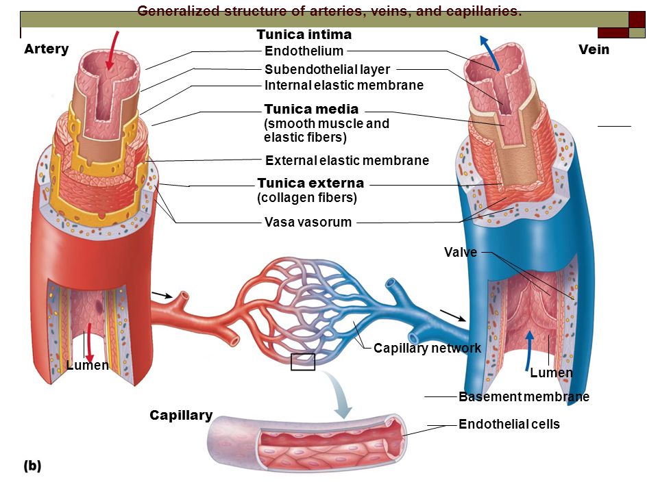
Arteries and arterioles have thicker walls than veins and venules because they are closer to the heart and receive blood that is surging at a far greater pressure ( Figure 20.3 ). Each type of vessel has a lumen —a hollow passageway through which blood flows.
Pictures Of Artery
Cross-section through valve Controlling blood flow In order to control blood flow through the vessels, the smooth muscle surrounding the arteries can constrict which causes vasoconstriction.
Arteries Histology Concise Medical Knowledge
Blood vessel histology Author: Lorenzo Crumbie MBBS, BSc • Reviewer: Dimitrios Mytilinaios MD, PhD Last reviewed: October 30, 2023 Reading time: 17 minutes It would be impossible to get blood to the predestined locations without the vascular pathways. Blood vessels form the extensive networks by which blood leaves the heart to supply tissue. . Additionally, other blood vessels return from.
Blood Vessels Alisa Houghton
Take a look at this cross-section through an elastic artery, and identify the three main layers - tunica intima, tunica media and tunica adventitia. Elastic arteries: These arteries that receive blood directly from the heart - the aorta and the pulmonary artery.: These need to be elastic because: They are relatively thin compared to their diameter.

Crosssectional view of an artery showing adventitia (outermost layer),... Download Scientific
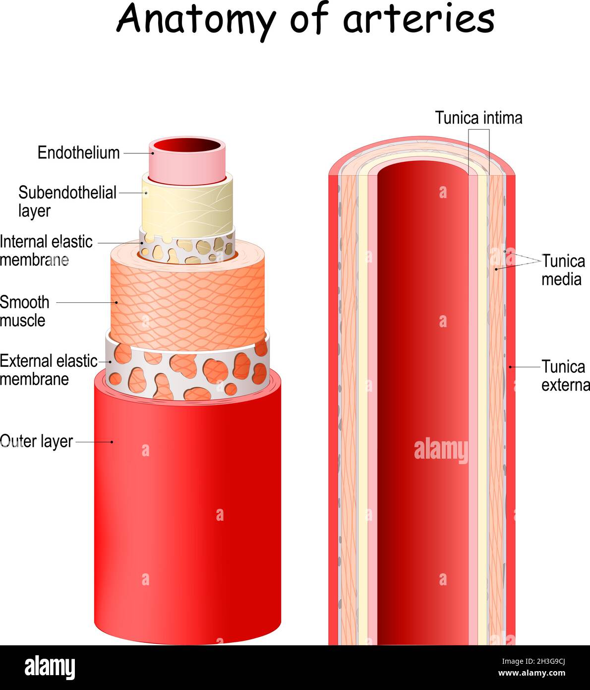
Structure Microscopic anatomy of an artery. Cross-section of a human artery The anatomy of arteries can be separated into gross anatomy, at the macroscopic level, and microanatomy, which must be studied with a microscope.

Artery crosssection Stock Image C007/9850 Science Photo Library
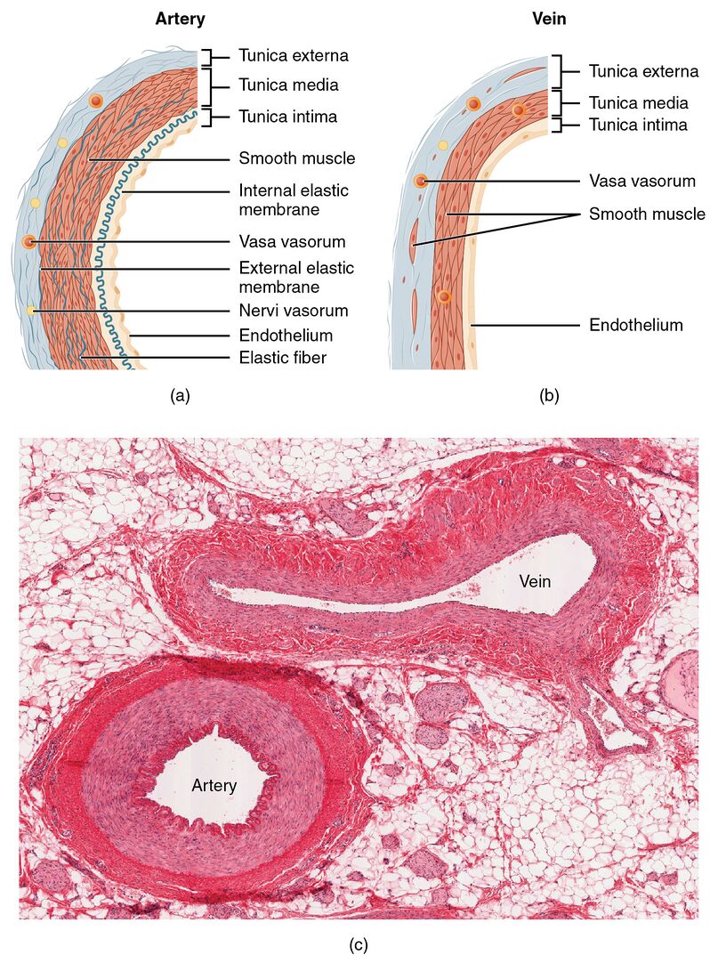
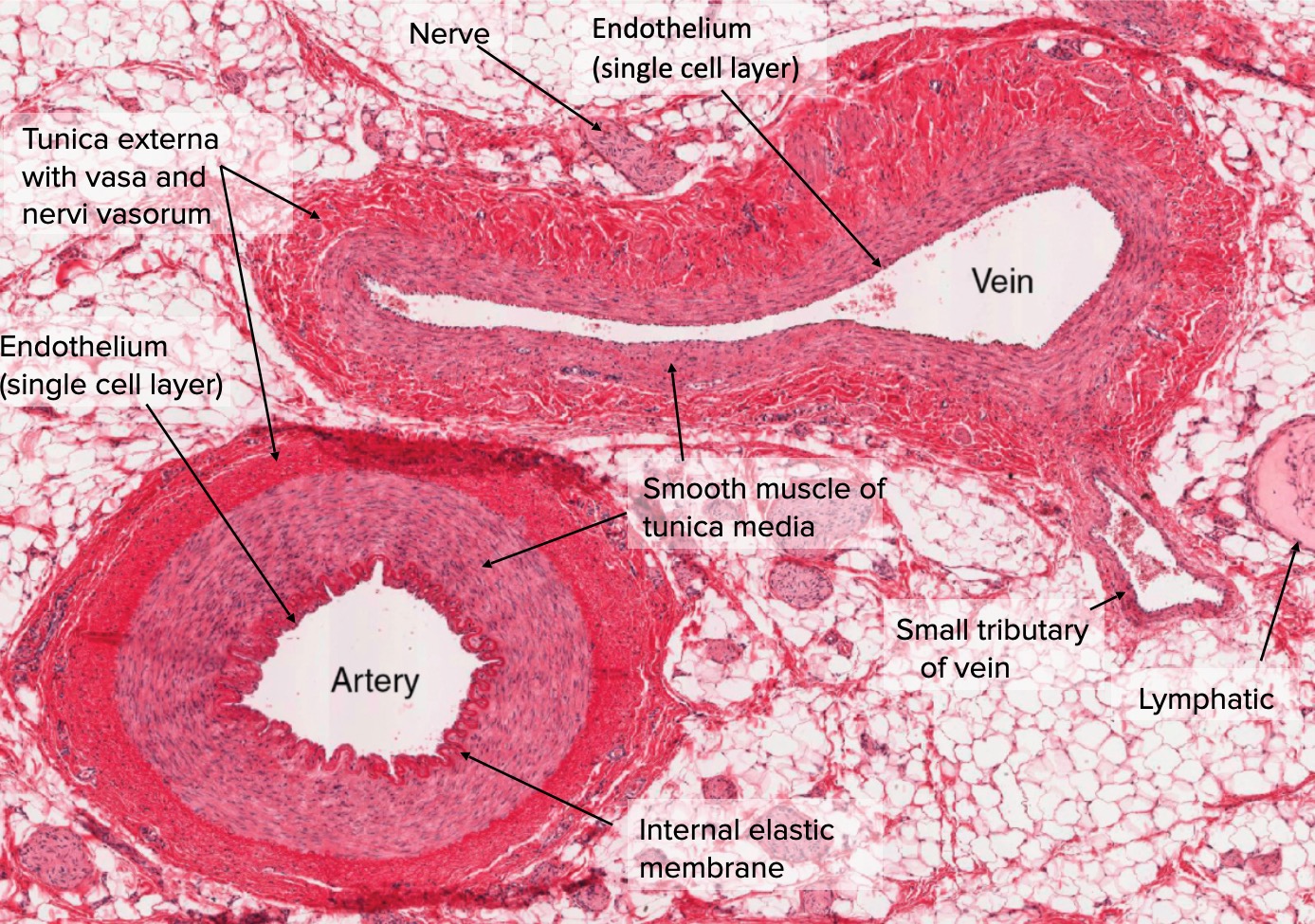
Arteries are tubular collections of cells which transport oxygenated blood and nutrients from the heart to the tissues of the body. Medical.. Cross section of artery and vein. Image: "Types of Arteries and Arterioles" by Phil Schatz. License: CC BY 4.0, edited by Lecturio.

Crosssectional views of the coronary artery. (A) Normal coronary... Download Scientific Diagram
Figure 40.10.1 40.10. 1: Blood vessel layers: Arteries and veins consist of three layers: an outer tunica externa, a middle tunica media, and an inner tunica intima. Capillaries consist of a single layer of epithelial cells, the endothelium tunic (tunica intima). Veins and arteries both have two further tunics that surround the endothelium: the.
Artery & Vein, cross section
Ventricular contraction ejects blood into the major arteries, resulting in flow from regions of higher pressure to regions of lower pressure, as blood encounters smaller arteries and arterioles, then capillaries, then the venules and veins of the venous system.. Figure 20.13 compares vessel diameter, total cross-sectional area, average blood.

Coronary Artery CrossSection Stock Image C004/8354 Science Photo Library
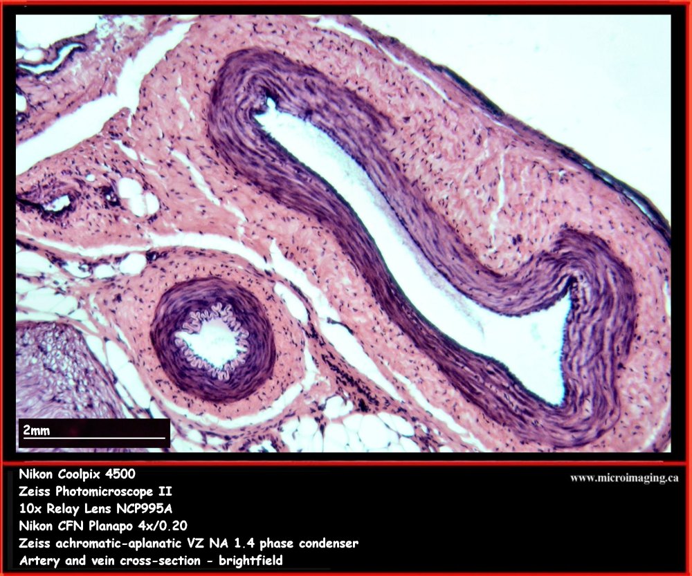
Arteries have smaller lumens than veins, a characteristic that helps to maintain the pressure of blood moving through the system. Together, their thicker walls and smaller diameters give arterial lumens a more rounded appearance in cross section than the lumens of veins. Figure \(\PageIndex{2}\): Structure of Blood Vessels.
Arteries vs Veins Structure, Function & Blood Flow
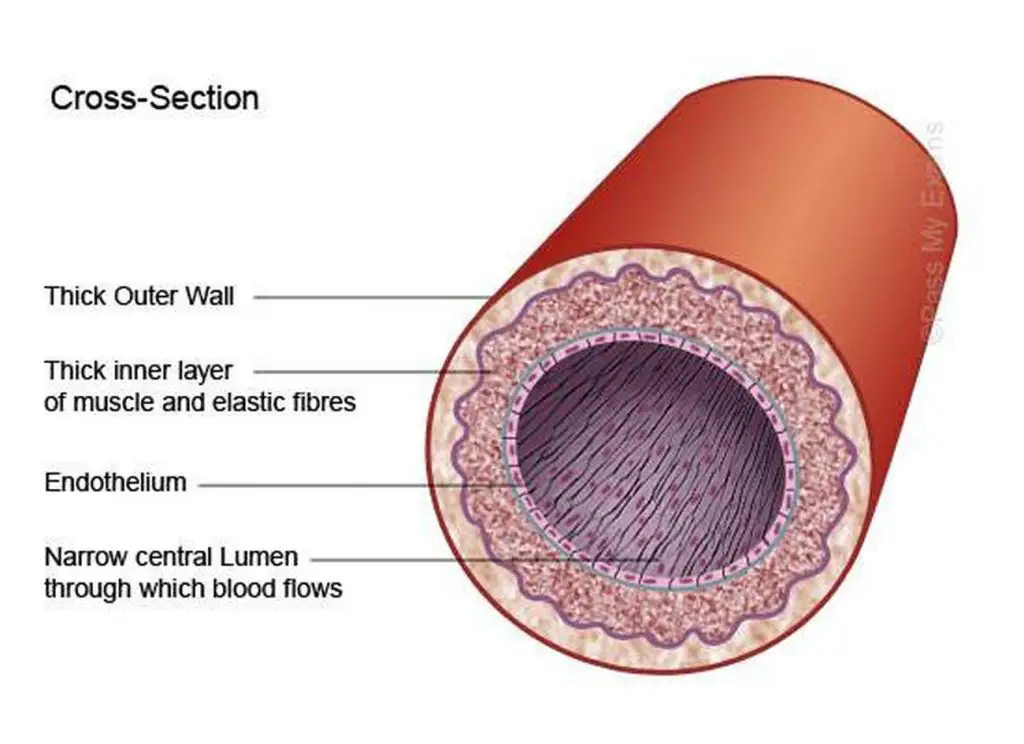
The lumen of an artery is shown in cross-section in the photomicrograph below. The width of blood vessels varies, but they all have a lumen. The walls of blood vessels differ depending on the type of vessel. In general, arteries and veins are more similar to one another than capillaries in the structure of their walls.

Cross Section Of An Artery
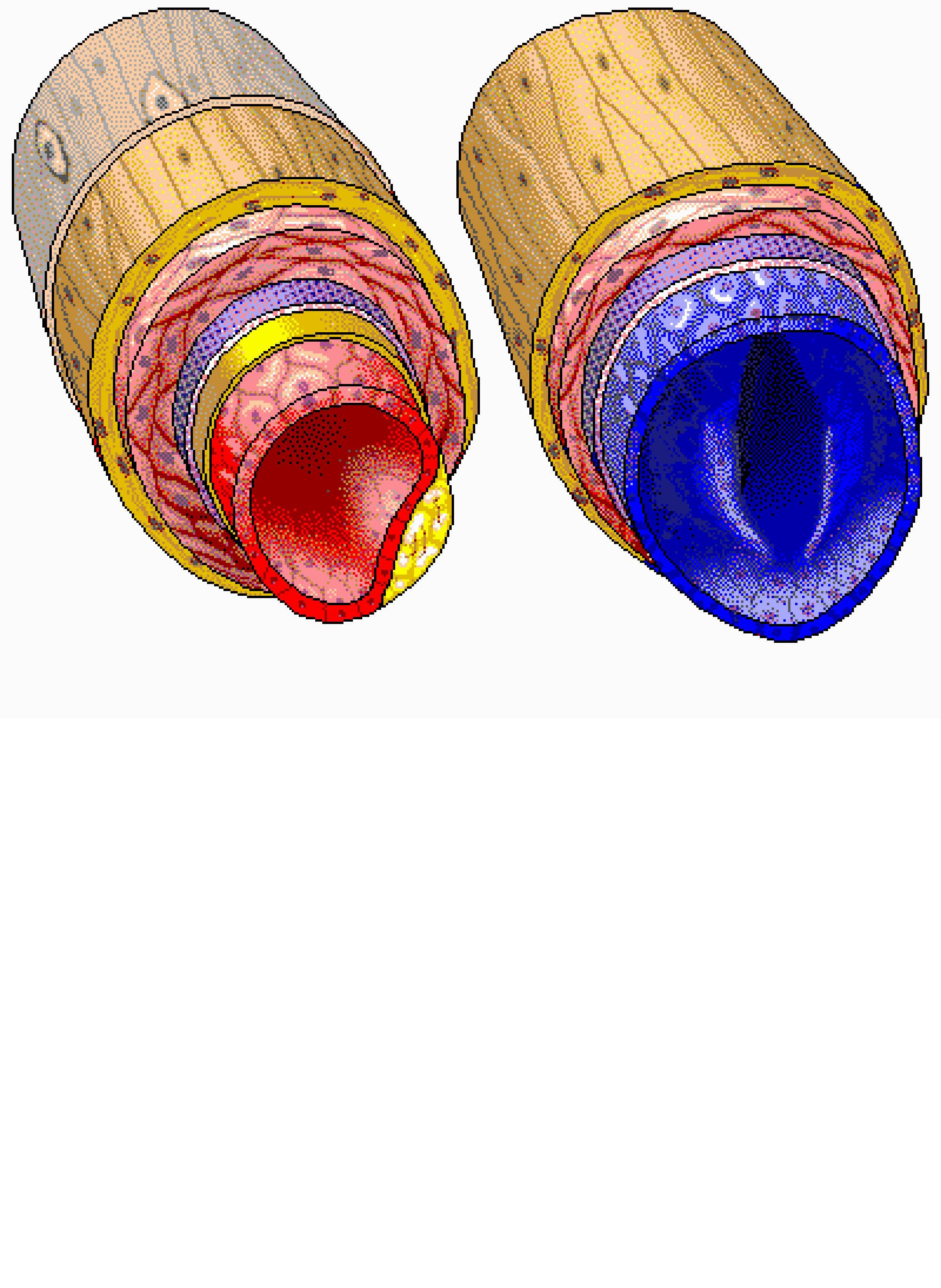
MedicalRF.com/Getty Images The artery wall consists of three layers: Tunica Adventitia (Externa) - the strong outer covering of arteries and veins. It is composed of connective tissue as well as collagen and elastic fibers.

Blood Vessel Structure and Function Boundless Anatomy and Physiology
96 Artery - Cross Section; Elastic Type Elastic Artery VIew Virtual EM Slide Elastic Artery. Note the alternating layers of connective tissue and smooth muscle cells in the media. If there are no fibroblasts in the media, which cell is involved in the synthesis and maintenance of the collagen and elastic fibers as well as vascular proteoglycans.
