Hand xray
visualizes the waist of scaphoid but is limited by overlap of carpal bones. scaphoid view. 30° wrist extension, 20° ulnar deviation. IR oblique. best view to see the waist and distal pole of scaphoid. if radiographs are negative and there is a high clinical suspicion, repeat radiographs in 14-21 days. Optional views.

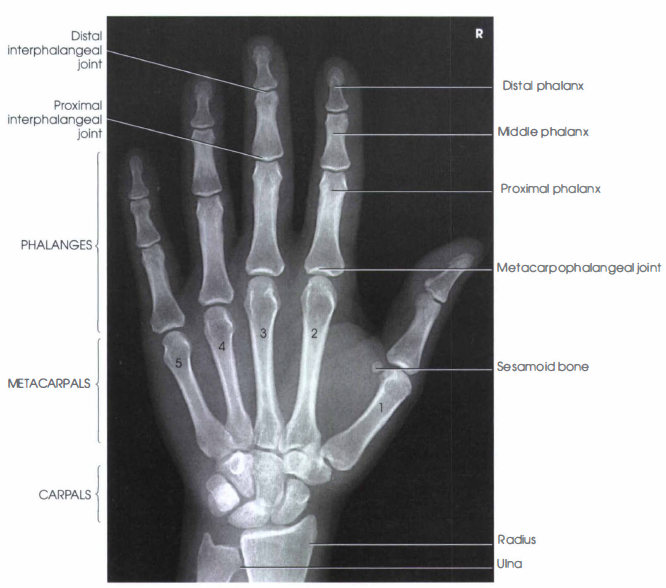
Hand Radiographic Anatomy wikiRadiography Radiology, Radiology student, Medical anatomy
Hand X-Ray Anatomy and Interpretation Checklist 1. Soft tissues - Look carefully at the soft tissue over all the bones for any swelling or foreign body. The swelling should prompt a careful search of the underlying bone or joint.⠀ 2. Bones - All the bones of the hand should be examined carefully and systematically.
Wrist Examination & Pathology Module Don't the Bubbles
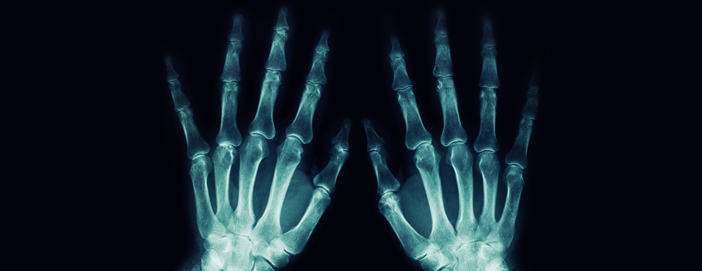
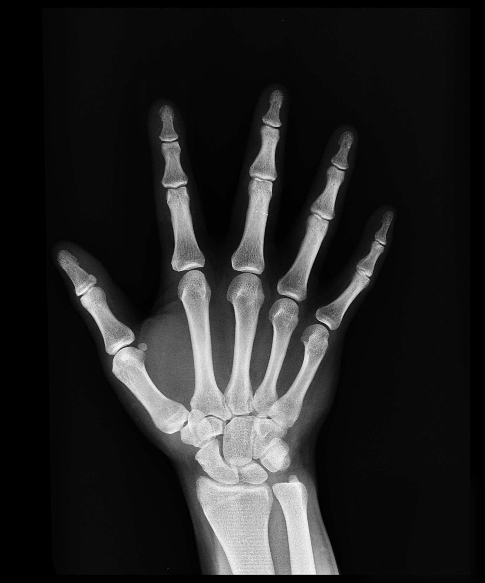
A hand X-ray (radiograph) is a test that creates a picture of the inside of your hand. The picture shows the inner structure ( anatomy) of your hand in black and white. Calcium in your bones absorbs more radiation, so your bones appear white on the X-ray.
Imaging Case of the Week 45 Emergucate
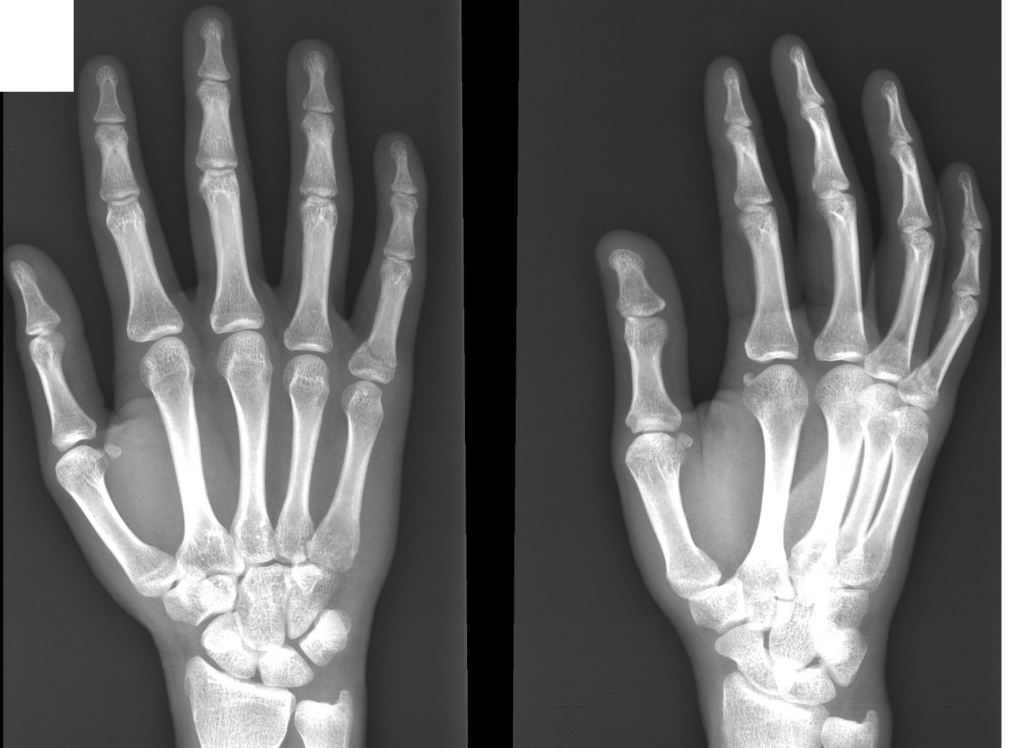
Summary indications suspicion of bony injury assessment of radiopaque foreign body assessment of joint disease procedure AP and oblique views of the hand a lateral view is of little use unless answering specific questions extends from the radiocarpal joint to the tips of fingers similar series wrist series

Read on to find out more about my review areas on a hand XRay... 👨🏽💻Hand xrays are
A hand X-ray is a black and white image that shows the inner structures of your hand, such as your bones and soft tissues. This diagnostic tool can help your doctor locate and understand injuries.

Wrist Radiographic Anatomy wikiRadiography Radiology student, Medical radiography, Medical
Zoe Little, specialty trainee 3 in trauma and orthopaedics, ; John Murphy, consultant orthopaedic surgeon; 1 Department of Trauma and Orthopaedics, Northwick Park Hospital, Harrow HA1 3UJ, UK; Correspondence to: zoe.little{at}doctors.org.uk

Hand xray. Causes, symptoms, treatment Hand xray
A recommended systematic checklist for reviewing musculoskeletal exams is: soft tissue areas, cortical margins, trabecular patterns, bony alignment, joint congruency, and review areas. Review the entire radiograph, regardless of perceived difficulty.
What a normal hand xray looks like (left) and what I managed to do to my hand(right). Local
Diagnostic Labels for Musculoskeletal Pain. Appropriate diagnosis of musculoskeletal pain involves a multifactorial approach that includes the history of the disease, a thorough physical.

Hand X Ray Medical Art Library
Key points. Finger injuries visible on X-ray include bone fractures, dislocations and avulsions. The hand comprises the metacarpal and phalangeal bones. Fractures and dislocations are usually straightforward to identify, so long as the potentially injured bone is fully visible in 2 planes. Finger joints commonly dislocate and are susceptible to.
Related Keywords & Suggestions for hand x ray anatomy
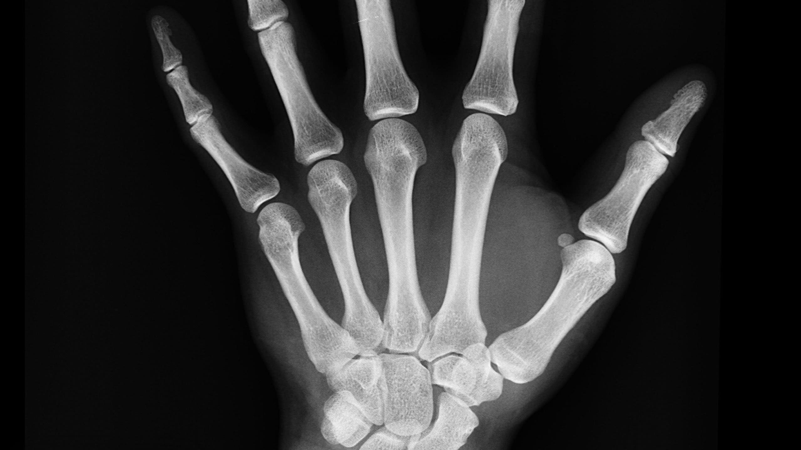
Hand x-rays are indicated for a variety of settings, including: trauma with suspected fracture suspected metacarpal dislocation foreign body detection and localization investigation of joint pain and/or deformity rheumatoid arthritis osteoarthrosis Projections Standard projections PA view

Normal Hand X Ray Colorvir Xray photo of normal right hand Stock Image Find the
Why the Test is Performed. Hand x-ray is used to detect fractures, tumors, foreign objects, or degenerative conditions of the hand. Hand x-rays may also be done to find out a child's "bone age." This can help determine if a health problem is preventing the child from growing properly or how much growth is left.
Hand XRay
The wrist is one of the most commonly requested X-Rays in the children's emergency department. Wrist views are requested when injury to the distal radius/ulna or carpal bones are suspected. Below is a systematic approach to interpretation.

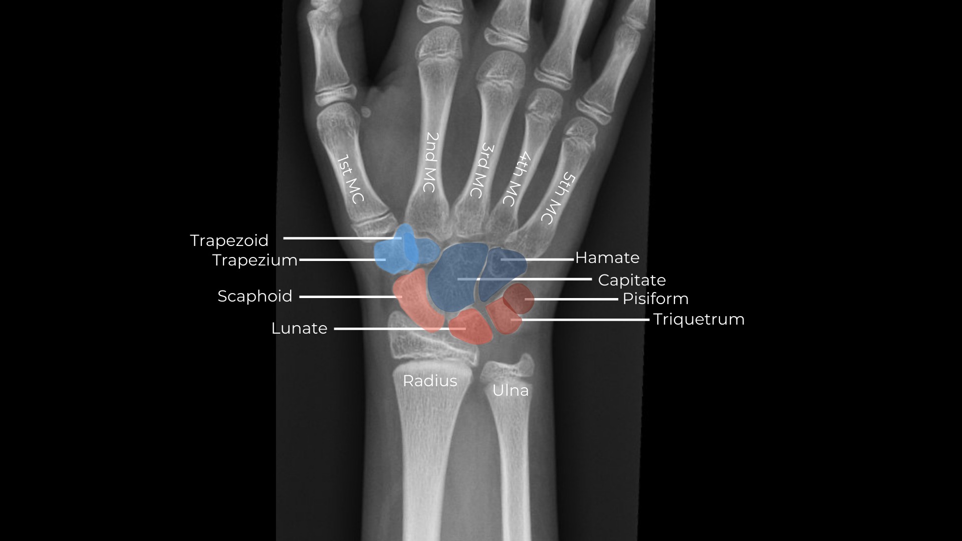
Xray image showing the left hand wrist in dorsal view. The carpal... Download Scientific Diagram
Wrist x-ray with labeled osseous anatomy. An official website of the United States government. Here's how you know.. [J Hand Surg Am. 2009] In vivo length changes of selected carpal ligaments during wrist radioulnar deviation. Xu J, Tang JB. J Hand Surg Am. 2009 Mar; 34(3):401-8.
5 ways to use Section Options
Labels: Base of fifth metacarpal Base of middle phalanx of middle finger Base of proximal phalanx of ring finger Capitate Distal phalanx of index finger Distal phalanx of thumb Hamate Head of fifth metacarpal Head of middle phalanx of middle finger Head of ulna Head of proximal phalanx of ring finger Hook of hamate Lunate Pisiform

Radiology Schools, Radiology Student, Radiology Technician, Radiology Imaging, Medical Imaging
This guide provides a step-by-step approach to interpreting wrist X-rays and includes examples of the key pathology you may come across. Anatomy The intricate anatomy of the wrist makes wrist X-ray interpretation a challenging task.

Frontal view x ray bones hand with Royalty Free Vector Image
Hand (oblique view) | Radiology Reference Article | Radiopaedia.org Hand (oblique view) Last revised by Andrew Murphy on 23 Mar 2023 Edit article Citation, DOI, disclosures and article data The hand oblique view is part of a two view series metacarpals, phalanges, carpal bones and distal radial ulnar joint. Indications
