
RxDentistry Radiographic Anatomy of Facial Bones
Appearances of facial bone fractures as seen on X-ray. X-ray of facial bones. McGriggor-Campbell fracture lines on facial bone x-rays. Use of OM views - occipitomental X-rays for diagnosis of facial bone fractures. The zygomatic arch looks like an elephants trunk on facial bone X-rays. Description of zygomatic arch fractures, trpod fractures and blow out fractures of the facial bones as seen.

2,358 X Ray Human Head Photos Free & RoyaltyFree Stock Photos from Dreamstime
Compare the injured side with the uninjured side. Fractures of the Facial Skeleton. Within the facial skeleton, there are relative areas of strength, which tend to be spared by fractures lines. These are: Alveolar ridge of the maxilla. Nasofrontal process of the maxilla. Body of the zygoma. Fractures of the Orbits.

Facial bone xrays
Written on 02/12/2016 , Last updated 31/07/2021 Cite this article as: Tessa Davis . Facial bone x-rays, Don't Forget the Bubbles, 2016. Available at: https://doi.org/10.31440/DFTB.10471 There are two views - occipito-mental view and occipito-mental 30 o view

XRay of Skull and Face Stock Image C039/4288 Science Photo Library
A facial X-ray is a series of pictures of the bones in the face. One type of facial X-ray (called a paranasal sinus X-ray series) looks at the air-filled cavities (sinuses) around the nose and eyes. A facial X-ray helps find bone fractures, tumours, foreign objects, infections, and abnormal growths or changes in bone structure or size.

Front face skull xray image Stock Photo Colourbox
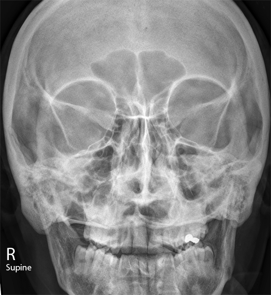
Cases and figures. Figure 1: cranial landmarks. Figure 2: skull positioning lines. Case 1: normal Waters view skull x-ray. Case 2: normal facial bones. Case 3: with zygomatic arch fracture. Case 4: orbital blowout fracture with teardrop sign. Case 5: maxillary and frontal sinusitis.
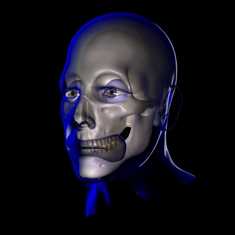
3d xray male face
A facial X-ray is a series of pictures of the bones in the face. One type of facial X-ray (called a paranasal sinus X-ray series) looks at the air-filled cavities (sinuses) around the nose and eyes. A facial X-ray helps find bone fractures, tumors, foreign objects, infections, and abnormal growths or changes in bone structure or size. An X-ray.

Facial Bone XRay Anatomy AP by Dr. Naveen Sharma GrepMed
Image receptor: 10 × 12 inch (24 × 30 cm) lengthwise. The reverse Waters method is used to show the facial bones when the patient cannot be placed in the prone position. Position of patient: • With the patient in the supine position, center the midsagittal plane of the body to the midline of the grid. Position of part:

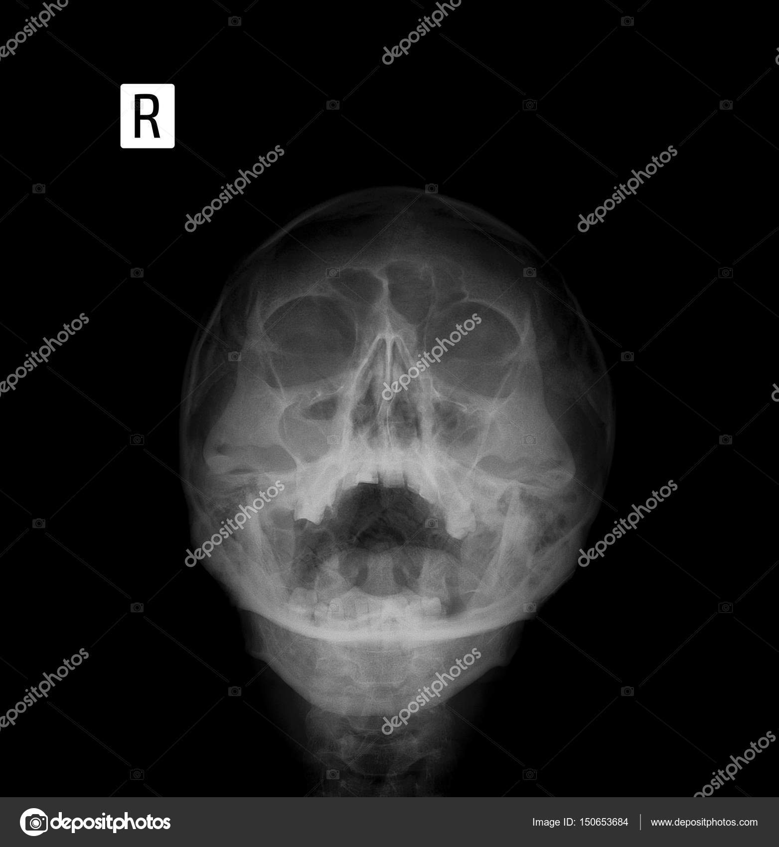
Xray/Face front stock photo. Image of diagnostic, illness 198832
A facial X-ray is a series of pictures of the bones in the face. One type of facial X-ray (called a paranasal sinus X-ray series) looks at the air-filled cavities (sinuses) around the nose and eyes. A facial X-ray helps find bone fractures, tumors, foreign objects, infections, and abnormal growths or changes in bone structure or size.
Xray film of the face frontal, nosechin projection. Sinusitis. — Stock Photo © vanzittoo
X-rays are a type of radiation that can pass through the body. They can't be seen by the naked eye and you can't feel them. As they pass through the body, the energy from X-rays is absorbed at different rates by different parts of the body.

Brace Face
Presentation Mild bruising and swelling over the right cheek after being assaulted, GCS of 15. Patient Data Age: 40 years Gender: Female x-ray Normal examination. No displaced facial or skull fracture noted. Case Discussion
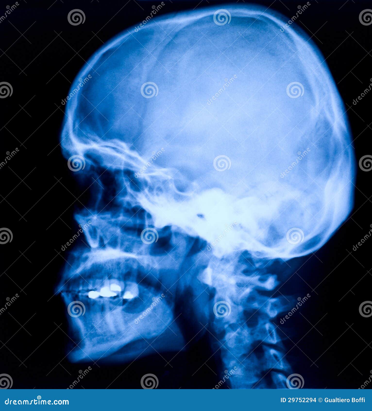
Head xray stock photo. Image of head, face, clinic, health 29752294
Traditionally, facial x-rays played an important screening role in the evaluation of facial trauma and infection. However, the complex three-dimensional relationship of facial bones and sinus air spaces makes interpretation of plain x-ray difficult.
Xray picturehuman head stock photo. Image of diagnosis 10611516
Unofficial implementation of paper 'Face X-ray for More General Face Forgery Detection'. (updating.) This is an unofficial implementation of Lingzhi Li, Jianmin Bao, Ting Zhang, Hao Yang, Dong Chen, Fang Wen, Baining Guo: Face X-Ray for More General Face Forgery Detection. CVPR 2020: 5000-5009.
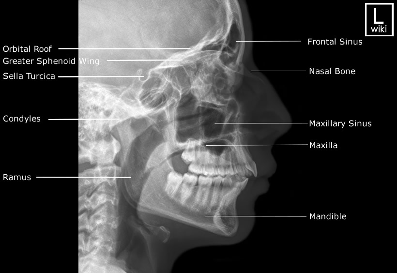
Facial Bones Radiographic Anatomy wikiRadiography
The zygomatic arch fracture is more easily seen on the OM30 (Occipito-Mental 30°) image. On the left (the non-injured side) overlying structures give the impression of a fracture, but careful scrutiny shows the cortex is intact. Description of zygomatic arch fractures of the face as seen on X-ray. Look for the 'elephant's trunk' appearance of.
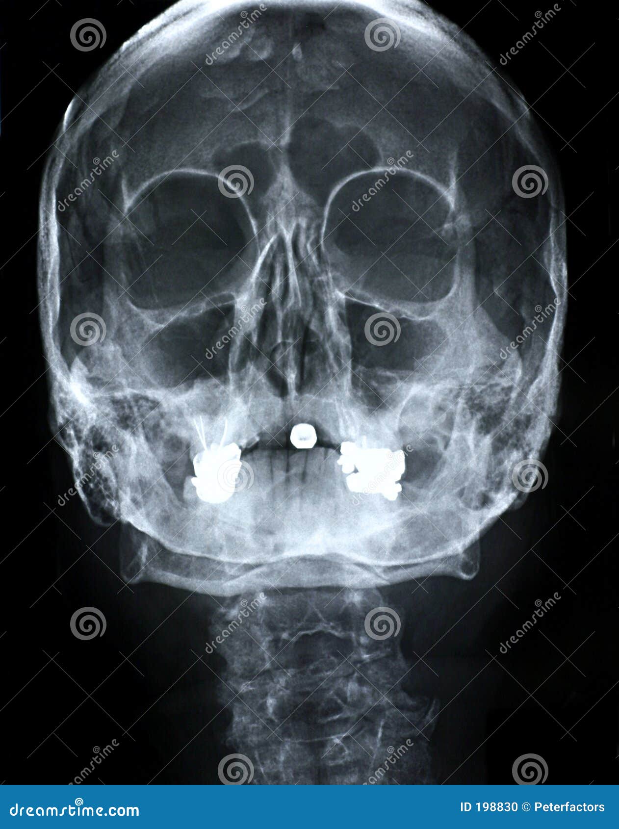
Xray/Face Front Stock Photo Image 198830
A skull X-ray is an imaging test doctors use to examine the bones of the skull, including the facial bones, the nose, and the sinuses. See a Body Map of the skull. It's an easy, quick, and.
Facial Bone Radiography wikiRadiography
Face X-ray is a method of radiation diagnosis of pathology of facial bones. It is used for visualization of the palatine, sublingual, maxillary, nasal, zygomatic bones and temporomandibular joint.

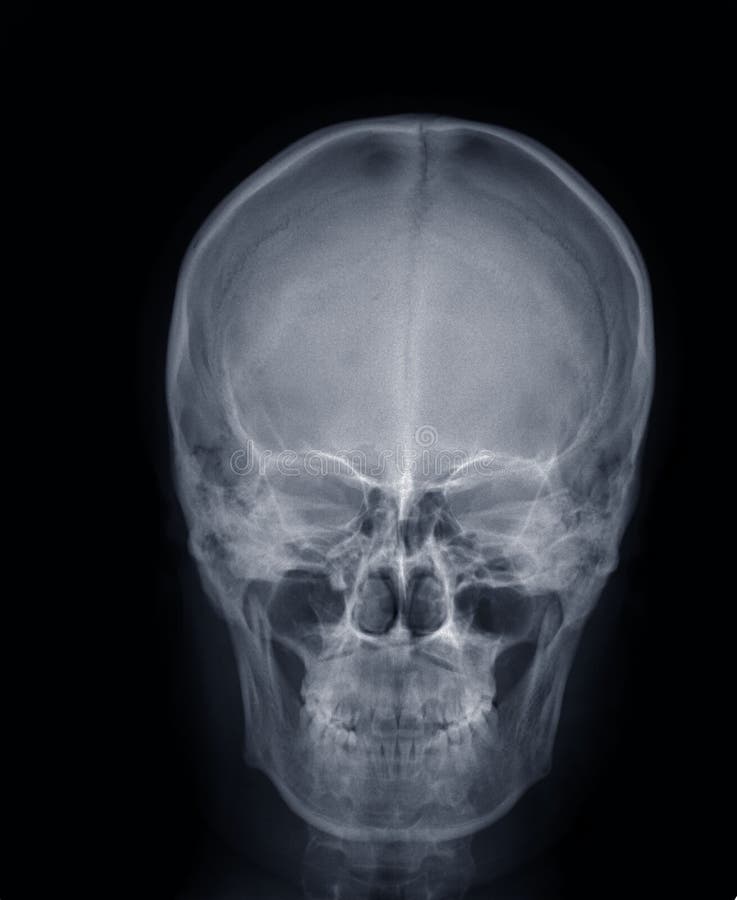
Normal Childs Head Xray Foto de stock Getty Images
A facial X-ray is a series of pictures of the bones in the face. One type of facial X-ray (called a paranasal sinus X-ray series) looks at the air-filled cavities (sinuses) around the nose and eyes. A facial X-ray helps find bone fractures, tumors, foreign objects, infections, and abnormal growths or changes in bone.
