Foot Anatomy Bones, Muscles, Tendons & Ligaments
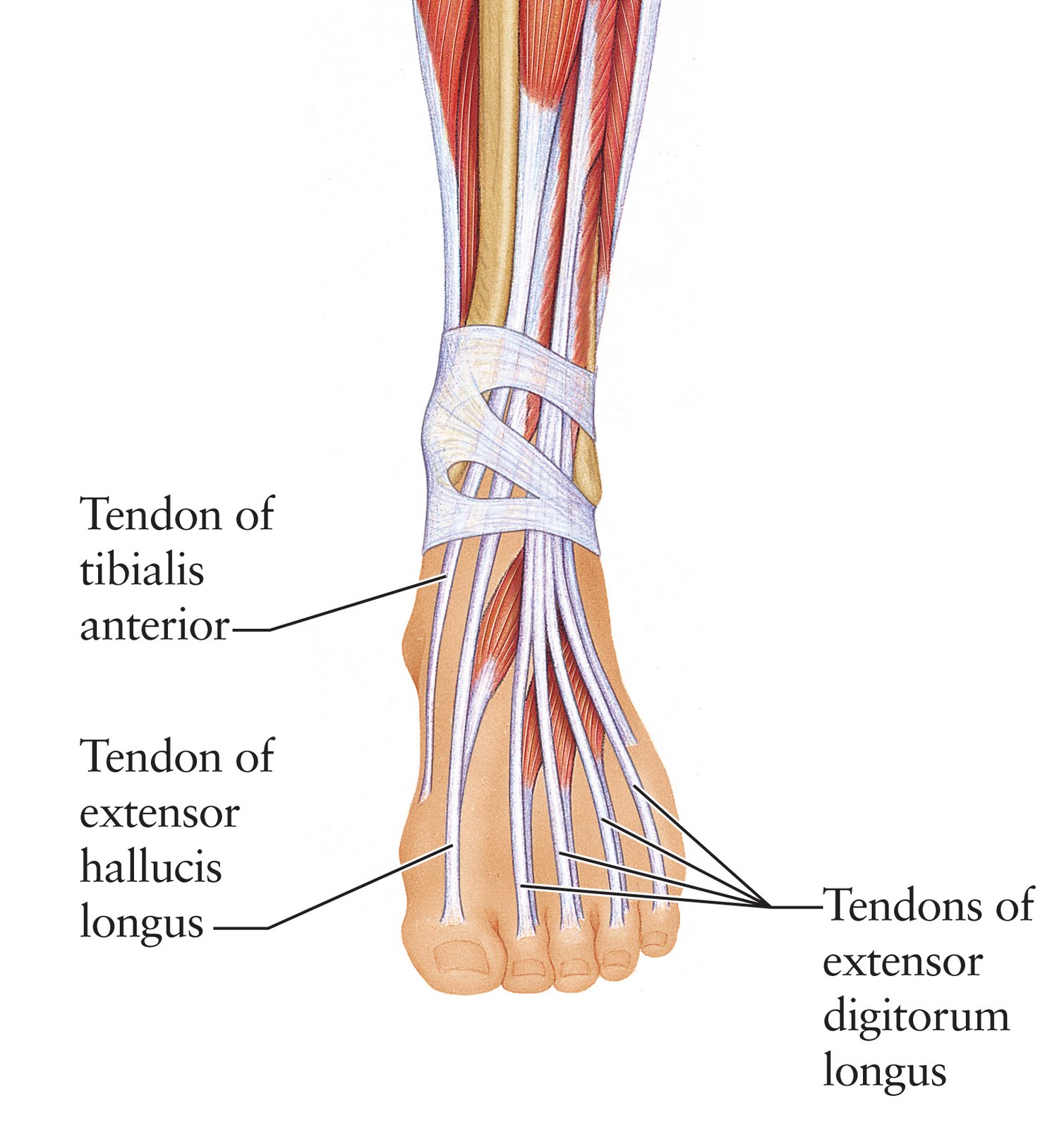
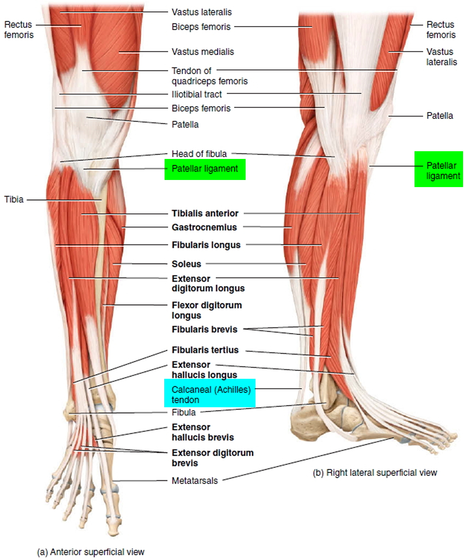
1. Tibialis Anterior Tendon The tibialis anterior muscle originates from the outer side of the tibia and passes down the front of the shin. The muscle turns into tendon about two thirds of the way down the shin and travels across the front of the ankle joint to the inner side of the foot underneath the medial foot arch.

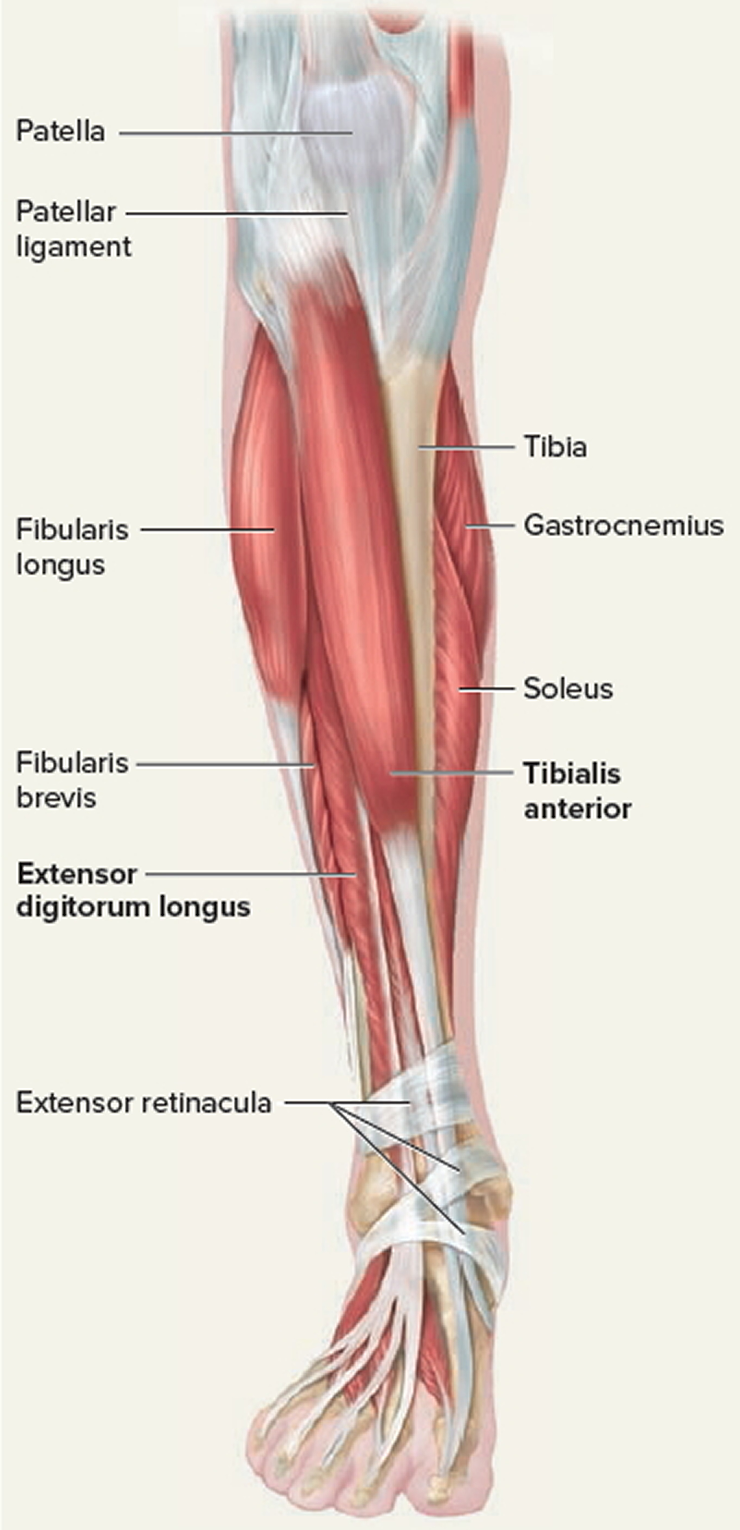
Tendon Diagram Leg muscles leg tendons hamstrings diagram
Structure Tendons are a type of dense, regular connective tissue. They are primarily made of strands of protein called collagen. These strands run parallel to each other to allow your tendons to effectively and efficiently transmit the force produced by your muscles to move your bones.
Tendon Function, Arm, Hand Tendons Leg and Achilles Tendons
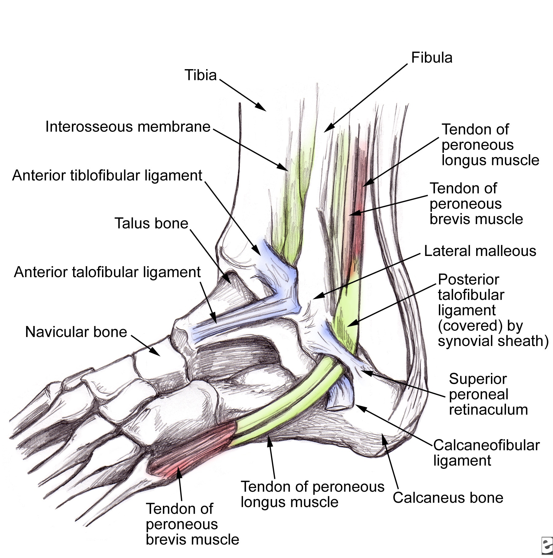
Ankle Joint. The ankle joint is where your shin bone (tibia), calf bone (fibula) and talus bone meet. It joins your foot to your lower leg. Your ankle also contains cartilage, ligaments, muscles, nerves and blood vessels. Your ankles move in two main directions and you use them any time you move your feet and legs.
Foot (Anatomy) Bones, Ligaments, Muscles, Tendons, Arches and Skin
33 joints more than 100 muscles, tendons, and ligaments Bones of the foot The bones in the foot make up nearly 25% of the total bones in the body, and they help the foot withstand weight..

image lateral_ankle for term side of card Ligament Tear, Ligaments And
The tendons in the foot are thick bands that connect muscles to bones. When the muscles tighten (contract) they pull on the tendons, which in turn move the bones. Arguably, the most important tendon is the Achilles tendon, which allows the calf muscles to move the ankle joint.

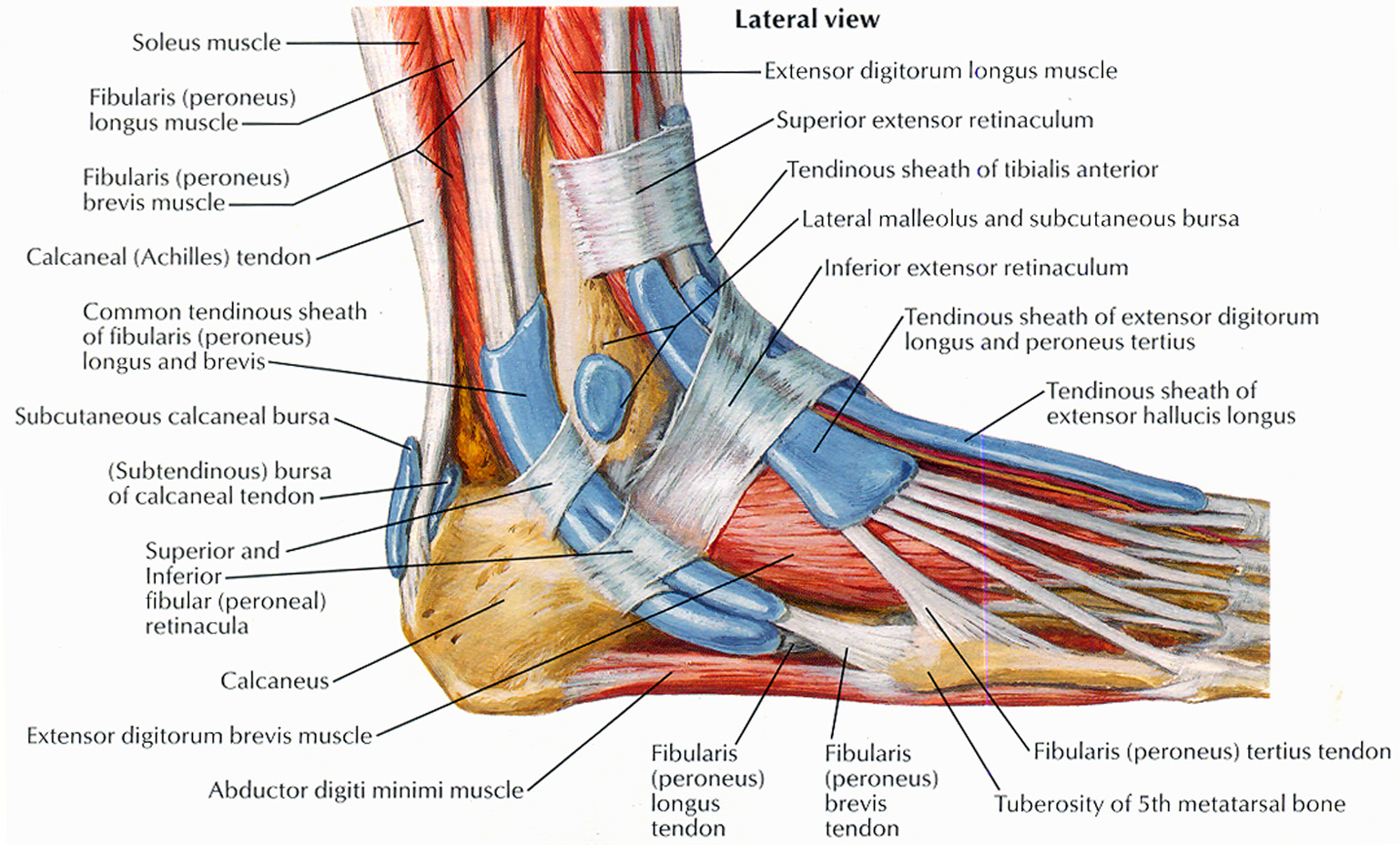
Tendons And Ligaments In Foot And Leg Lateral Ankle Anatomy Lower Leg
Forward movement (propulsion) The foot must be flexible to adapt to uneven surfaces and remain stable when you're walking. The foot has three parts: the forefoot, midfoot, and hindfoot. There are bones, joints, muscles, tendons, and ligaments in each of these sections. Orientation of the Foot The bottom part of the foot is the sole.
Human Anatomy for the Artist The Dorsal Foot How Do I Love Thee? Let
Tendons in the Foot Diagram: Understanding the Anatomy and Function. The foot is a complex structure composed of bones, muscles, ligaments, and tendons. While all these components work together to support our body weight and facilitate movement, it's the tendons in the foot that play a crucial role in transmitting the force generated by the.

Tendon Diagram / Foot Ankle Tendonitis Causes Symptoms Treatment In
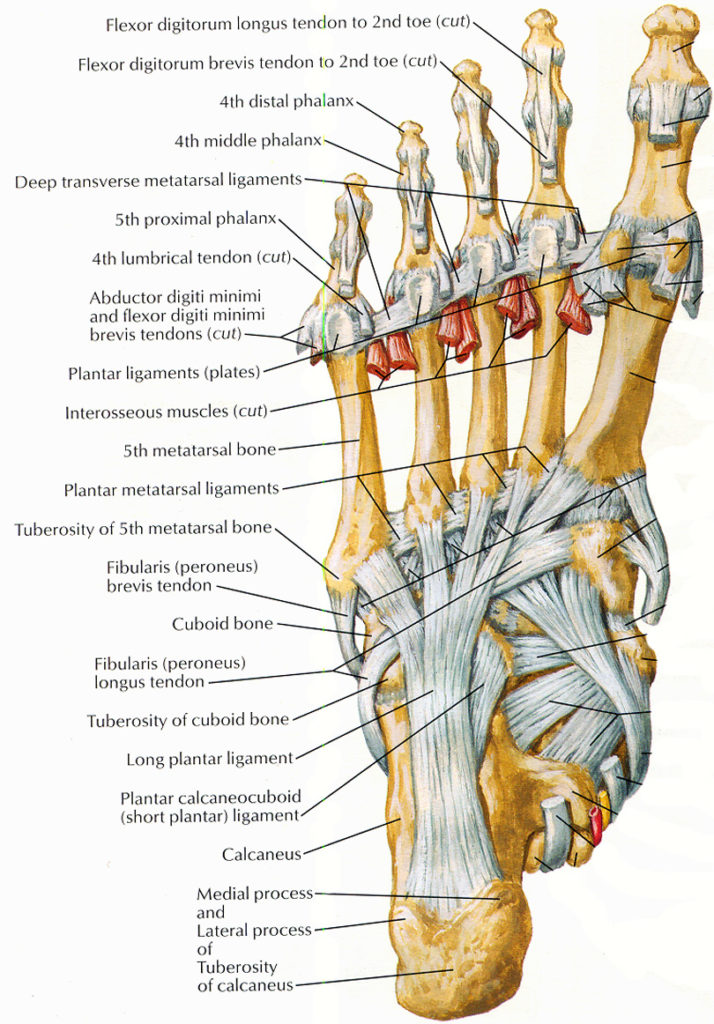
Foot Ligaments Foot Ligaments Your feet are complex and hard-working body parts. They contain 26 bones, 30 joints and over 100 muscles, tendons and ligaments. Your foot includes three main ligaments that connect your bones and provide support for the arch of the foot.

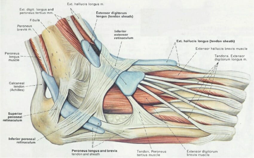
Diagram showing the tendons and ligaments of the ankle and foot
4 Main Motions of the Foot Dorsal flexion (pulling the foot and toes upward): The main tendon for this movement is the Anterior Tibialis. Plantar flexion (pointing the foot and toes downward): The main tendon for this is the Achilles Tendon. Inversion (turning the foot inward): The main tendon for this is Posterior Tibialis Tendon.
ligaments and tendons of foot netter CoreWalking
The foot's complex structure contains more than 100 tendons, ligaments, and muscles that move nearly three dozen joints, while bones provide structure.
Strained Peroneal Tendon...? run.around.aroo
Overview What is foot tendonitis? Foot tendonitis (tendinitis) is inflammation or irritation of a tendon in your foot. Tendons are strong bands of tissue that connect muscles to bones. Overuse usually causes foot tendonitis, but it can also be the result of an injury. Are there different types of foot tendonitis? Your feet contain many tendons.
Human Anatomy for the Artist The Dorsal Foot How Do I Love Thee? Let
There are a variety of anatomical structures that make up the anatomy of the foot and ankle (Figure 1) including bones, joints, ligaments, muscles, tendons, and nerves. These will be reviewed in the sections of this chapter. Figure 1: Bones of the Foot and Ankle Regions of the Foot
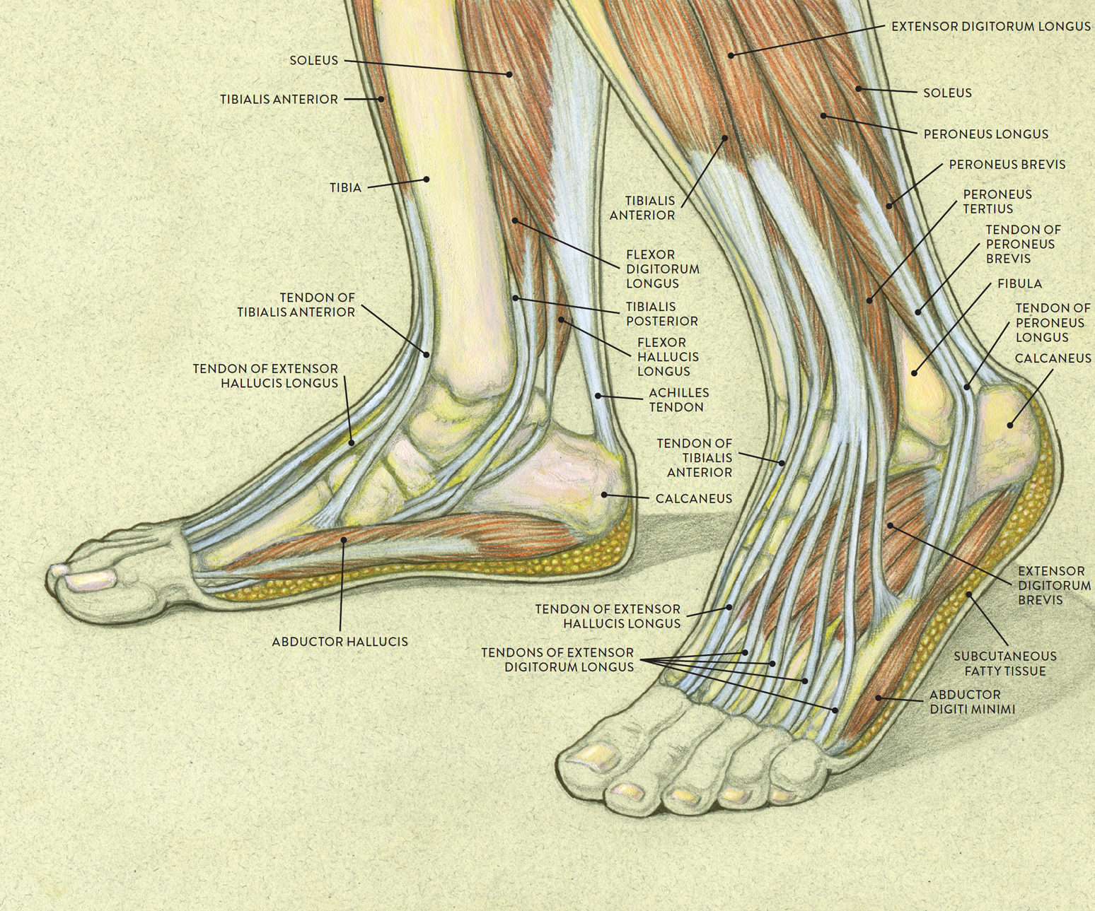
Muscles of the Leg and Foot Classic Human Anatomy in Motion The
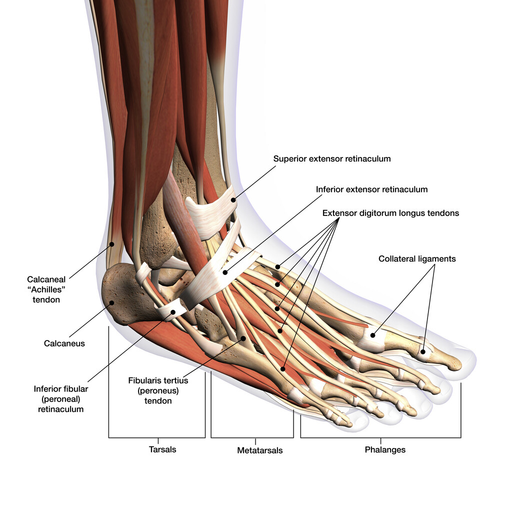
Ankle joint Explore study unit Joints and ligaments of the foot Explore study unit Bones of the foot There are 26 bones in the foot, divided into three groups: Seven tarsal bones Five metatarsal bones Fourteen phalanges Tarsals make up a strong weight bearing platform.
Tendons of the Foot JOI Jacksonville Orthopaedic Institute
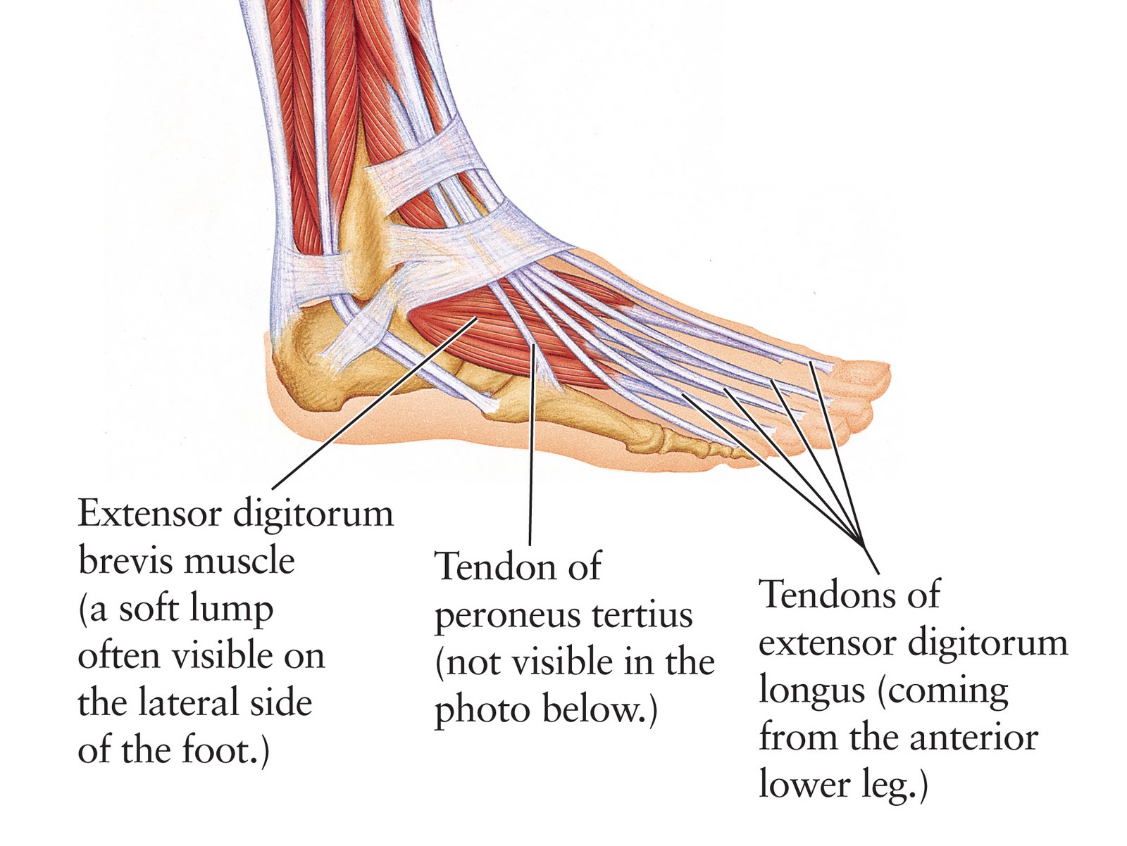
The extensor digitorum brevis is a small, thin muscle which lies underneath the long extensor tendons of the foot. Attachments: Originates from the calcaneus and inferior extensor retinaculum. It attaches onto the long extensor tendons of the medial four toes. Actions: Extension of the lateral four toes.

Foot Description, Drawings, Bones, & Facts Britannica
This tendon in the back of the calf and ankle connects the plantaris, calf, and soleus muscles to the heel bone. It stores the elastic energy needed for running, jumping, and other physical.
Tendinopathy causes, symptoms, diagnosis, treatment and exercises
1/2 Synonyms: Talocrural joint The foot is the region of the body distal to the leg that is involved in weight bearing and locomotion. It consists of 28 bones, which can be divided functionally into three groups, referred to as the tarsus, metatarsus and phalanges.
